. 2008 Apr;29(10):1454-63.
doi: 10.1016/j.biomaterials.2007.11.041. Epub 2007 Dec 27.

Small-diameter biodegradable scaffolds for functional vascular tissue engineering in the mouse model

Affiliations

Small-diameter biodegradable scaffolds for functional vascular tissue engineering in the mouse model

Jason D Roh et al. Biomaterials. 2008 Apr.

Abstract

The development of neotissue in tissue engineered vascular grafts remains poorly understood. Advances in mouse genetic models have been highly informative in the study of vascular biology, but have been inaccessible to vascular tissue engineers due to technical limitations on the use of mouse recipients. To this end, we have developed a method for constructing sub-1mm internal diameter (ID) biodegradable scaffolds utilizing a dual cylinder chamber molding system and a hybrid polyester sealant scaled for use in a mouse model. Scaffolds constructed from either polyglycolic acid or poly-l-lactic acid nonwoven felts demonstrated sufficient porosity, biomechanical profile, and biocompatibility to function as vascular grafts. The scaffolds implanted as either inferior vena cava or aortic interposition grafts in SCID/bg mice demonstrated excellent patency without evidence of thromboembolic complications or aneurysm formation. A foreign body immune response was observed with marked macrophage infiltration and giant cell formation by post-operative week 3. Organized vascular neotissue, consisting of endothelialization, medial generation, and collagen deposition, was evident within the internal lumen of the scaffolds by post-operative week 6. These results present the ability to create sub-1mm ID biodegradable tubular scaffolds that are functional as vascular grafts, and provide an experimental approach for the study of vascular tissue engineering using mouse models.


Figures
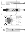
Fig. 1
Fig. 1
Schematic of scaffold fabrication using the dual cylinder chamber system. A) Flat polyester felts were easily rolled into tubular constructs using a gradually tapered cylinder. B) P(CL/LA) solution sealed the nonwoven polyester tube by creating an interconnecting porous structure between the felt fibers. C) Cross sectional image demonstrating the configuration of the components and removal of solvents by lyophilization. D) Resultant hybrid scaffold pushed out through inlet after inner needle cylinder removed.
Fig. 2
Fig. 2
Structural characterization of scaffolds. A) SEM of PGA-P(CL/LA) scaffold (0.9mm I.D., 150µm wall thickness). B) SEM of PLLA-P(CL/LA) scaffold (0.7 mm I.D., 250µm wall thickness). C) Pore size distribution comparing uncoated nonwoven PGA felt to PGA-P(CL/LA) scaffold. D) Pore size distribution comparing uncoated nonwoven PLLA felt to PLLA-P(CL/LA) scaffold.
Fig. 3
Fig. 3
Biomechanical characterization of scaffolds. A) Burst pressure. B) Suture retention strength. C) Young’s modulus. D) Tensile strength of PGA-P(CL/LA) and PLLA-P(CL/LA) scaffolds over 24wk time course.
Fig. 4
Fig. 4
Scaffolds biocompatible with human cell seeding. Scanning electron photomicrographs of: A) Unseeded PGA-P(CL/LA) scaffold, B) Unseeded PLLA-P(CL/LA) scaffold, C) PGA-P(CL/LA) scaffold seeded with hAoSMC, and D) PLLA-P(CL/LA) scaffold seeded with hAoSMC. Hematoxylin and eosin stains of E) PGA-P(CL/LA) scaffold seeded with hAoSMC and F) PLLA-P(CL/LA) scaffold seeded with hAoSMC. G) MTT assay comparing hAoSMC viability on PGA-P(CL/LA), PLLA-P(CL/LA), and polystyrene shows no significant differences. Original magnification 200X (E, F).
Fig. 5
Fig. 5
Scaffolds elicit foreign body reaction with giant cell formation (O) by postoperative wk 3 when implanted as vascular interposition grafts. Representative images of PGA-P(CL/LA) venous grafts stained with A) H&E (inner lumen), B) Mac-3 (inner lumen, brown=macrophage), C) Gomori trichrome (inner lumen, blue=collagen), and D) Gomori trichrome (outer lumen) are shown. Representative images of PLLA-P(CL/LA) arterial grafts stained with E) H&E (inner lumen), F) Mac-3 (inner lumen), G) Gomori trichrome (inner lumen), and H) Gomori trichrome (outer lumen) are shown. Original magnification 400X.
Fig. 6
Fig. 6
Scaffolds functioning as vascular grafts at post-operative wk 3. A) MicroCT reconstruction of PGA-P(CL/LA) scaffold as IVC graft, B) MicroCT reconstruction of PLLA-P(CL/LA) scaffold as aortic graft. By 3 wks, scaffolds are fully infiltrated with cells. C) H&E of PGA-P(CL/LA) as IVC graft. D) H&E of PLLA-P(CL/LA) as aortic graft. Original magnification 100X (C,D).
Fig. 7
Fig. 7
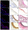
Development of organized vascular neotissue by post-operative wk 6. Representative images of PGA-P(CL/LA) venous graft stained with A) vWF (endothelial layer), B) αSMA (medial layer), and C) Verhoeff-van Gieson (black=elastin, pink=collagen). Representative images of native mouse IVC stained with D) vWF, E) αSMA, F) Verhoeff-van Gieson. Representative images of PLLA-P(CL/LA) arterial graft stained with G) vWF, H) αSMA, and I) Verhoeff-van Gieson. Representative images of native mouse aorta stained with J) vWF, K) αSMA, and L) Verhoeff-van Gieson. Original magnification 400X.

References

    1. Niklason LE, Gao J, Abbott WM, Hirschi KK, Houser S, Marini R, Langer R. Functional arteries grown in vivo. Science. 1999;284:489–493. - PubMed
    1. Kaushal S, Amiel GE, Guleserian KJ, Shapira OM, Perry T, Sutherland FW, Rabkin E, Moran AM, Schoen FJ, Atala A, Soker S, Bischoff J, Mayer JE. Functional small-diameter neovessels created using endothelial progenitor cells expanded ex vivo. Nat Med. 2001;7:1035–1040. - PMC - PubMed
    1. L’Heureux N, Dusserre N, Konig G, Victor B, Keire P, Wight TN, Chronos NA, Kyles AE, Gregory CR, Hoyt G, Robbins RC, McAllister TN. Human tissue-engineered blood vessels for adult arterial revascularization. Nat Med. 2006;12:361–365. - PMC - PubMed
    1. Watanabe M, Shin’oka T, Tohyama S, Hibino N, Konuma T, Matsumura G, Kosaka Y, Ishida T, Imai Y, Yamakawa M, Ikada Y, Morita S. Tissue engineered vascular autograft: inferior vena cava replacement in a dog model. Tissue Eng. 2001;7:429–439. - PubMed
    1. Campbell JH, Efendy JL, Campbell GR. Novel vascular graft grown within recipient’s own peritoneal cavity. Circ Res. 1999;85:1173–1178. - PubMed

Publication types