Review
. 2008 Jan;33(1):9-19.
doi: 10.1016/j.tibs.2007.09.014. Epub 2007 Dec 31.

Bacterial chemoreceptors: high-performance signaling in networked arrays

Affiliations
Review

Bacterial chemoreceptors: high-performance signaling in networked arrays

Gerald L Hazelbauer et al. Trends Biochem Sci. 2008 Jan.

Abstract

Chemoreceptors are crucial components in the bacterial sensory systems that mediate chemotaxis. Chemotactic responses exhibit exquisite sensitivity, extensive dynamic range and precise adaptation. The mechanisms that mediate these high-performance functions involve not only actions of individual proteins but also interactions among clusters of components, localized in extensive patches of thousands of molecules. Recently, these patches have been imaged in native cells, important features of chemoreceptor structure and on-off switching have been identified, and new insights have been gained into the structural basis and functional consequences of higher order interactions among sensory components. These new data suggest multiple levels of molecular interactions, each of which contribute specific functional features and together create a sophisticated signaling device.


Figures
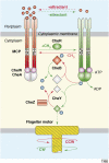
Figure I
Figure I
The chemoreceptor signaling pathway in E. coli. Components and reactions in red promote counter clockwise (CCW) flagellar rotation; those in green promote clockwise (CW) flagellar rotation. Components in gray represent inactive forms. Solid lines represent enzymatic reactions; broken lines indicate binding interactions. CheA-derived phosphoryl groups are shown as blue spheres. Receptor modification sites are shown as white (unmethylated) and black (methylated) circles.
Figure 1
Figure 1
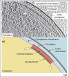
A patch of membrane-embedded chemoreceptors in a signaling complex near the pole of an intact cell. The emerging technique of cryo-electron tomography can provide images of the detailed internal structure of intact cells, including macromolecular complexes, without the need for fixation, staining or other potentially perturbing treatments. Application of this technique to motile and chemotactic E. coli cells [20] reveals distinct patches of striations, usually near a pole, extending perpendicular to the cytoplasmic membrane. These patches occur in cells containing chemoreceptors, the histidine kinase CheA and the coupling protein CheW, but not in cells lacking any one of these proteins. Immuno-electron microscopy demonstrates that the striations consist of chemoreceptors and that a thin line of density parallel to the membrane at the membrane-distal end of the striations contains CheA and CheW. (a) A 5 nm tomographic slice of the region near a pole of an intact, chemotactically wild-type E. coli cell [20]. The slice is oriented essentially normal to the two membranes (indicated by labeled arrows) that surround this Gram-negative cell – the cytoplasmic membrane, which is the cell's permeability barrier, and the outer membrane, which is a penetration barrier that creates the periplasm between the two membranes. A patch of chemoreceptors, CheA and CheW is visible along part of the cytoplasmic membrane. The boundaries of the patch are marked by white arrows. (b) Schematic of the tomograph in (a) showing the membranes (outer membrane, dark gray; cytoplasmic membrane, light gray), periplasm (light blue) and cytoplasm (yellow). The boundaries of the patch are marked by white arrows as in part (a). Chemoreceptors (red) and the layer of CheA and CheW (blue) are indicated by labeled arrows. Panel (a) is derived from the same original image from which Figure 2a of Zhang et al. [20] was made. We thank S. Subramaniam for that original image. Part (b) is based on Figure 1c of the same paper. Adapted, with permission, from [20].
Figure I
Figure I
Diagnostic cross-linking sites for the trimer-of-dimer organization. (a) Backbone traces of receptor signaling tips in a mixed trimer-of-dimers containing one Tsr molecule (yellow) and two Tar molecules (light blue). Residues chosen for cysteine reporter replacements: V384 (white, Tar), V398 (black, Tsr), S366 (blue, Tar and Tsr). (b) View down the trimer axis looking towards the tip. Note the trigonal arrangement of residues at position 366 in the inward-facing subunits of the dimers. (c) View of the trimer tip looking towards the membrane. Note the proximity of Tar residue 384 and Tsr residue 398 at a dimer–dimer interface in the trimer.
Figure 2
Figure 2
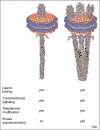
Functional activities of Nanodisc-embedded chemoreceptors. Nanodiscs are soluble, nanoscale (~10 nm diameter) particles of lipid bilayer surrounded by an annulus of amphiphilic membrane scaffold protein [39]. They form spontaneously when detergent is removed from mixtures of detergent-solubilized lipid and scaffold protein. If the preparation mixture contains detergent-solubilized membrane protein, the protein is incorporated into Nanodiscs. A Nanodisc-embedded protein is in a lipid bilayer and thus probably exists in its native state, but it is segregated from other membrane proteins. Chemoreceptors can be incorporated into Nanodiscs [40]. Preparations with different average number of chemoreceptor dimers per disc can be prepared by manipulating the input ratio of receptor to scaffold protein. These Nanodisc preparations enabled determination of chemoreceptor activity as a function of the number of receptor dimers per disc, that is the number of dimers that could potentially interact. The figure (a modified version of Figure 4 from Boldog et al. [40]), presents schematics of Nanodisc-embedded chemoreceptors in the onedimer per disc (left) or three-dimers per disc (right) state (shown as a trimer-of-dimers) and indicates the receptor activities exhibited in the two different conditions. Preparations of one dimer per Nanodisc were effectively methylated, and initial rates of this modification were doubled by the presence of a saturating concentration of an attractant ligand. These observations indicated that individual chemoreceptor dimers reconstituted in Nanodiscs were substrates for adaptational modification and coupled ligand-binding to methylation propensity. However, preparations of one chemoreceptor dimer per disc were ineffective at activating the chemotaxis kinase CheA. Nanodiscs with three dimers per disc exhibited a relatively sharp maximum of kinase activation, and activation was eliminated by the addition of a saturating concentration of an attractant ligand [40]. These observations indicated that functionally relevant kinase activation required the combined action of more than one chemoreceptor and were consistent with the functional importance of three dimers, for instance as a trimer-of-dimers. Adapted, with permission, from [40].
Figure 3
Figure 3
Conformational signaling in chemoreceptor dimers. The left-hand schematic, labeled ’Attractant response’ shows the conformational changes that convey an informational signal from one end of the chemoreceptor to the other. The right-hand schematic, labeled ’Sensory adaptation’, shows how covalent modification of an attractant-occupied receptor (methylation of specific glutamyl residues) reverses the ligand-induced conformational changes and thus mediates sensory adaptation. For clarity, a single receptor dimer is shown, embedded in the cytoplasmic membrane (light gray rectangle). As seen in the left-hand schematic, binding of an attractant chemoeffector (gray circle) to the transmembrane sensing module (green) initiates downward displacement of the signaling helix (see arrow heads on the signaling helix of the transmembrane sensing module) [49]. This movement generates an as-yet undefined conformational change in the signal conversion module (gray), which shifts its signaling state. This shift in turn weakens subunit interactions (symbolized by heavy arrows pointing away from the interface of subunit interaction) in the kinase control module (blue), resulting in increased flexibility, twisting and/or bending of the receptor molecule [,–58]. These changes, either directly or through effects on the trimer [47,48,60], deactivate coupled CheA kinase molecules leading to a counter-clockwise motor response. The same conformational changes also increase the propensity of the kinase control module for methylation and decrease its propensity for demethylation. The ligand-induced kinase inhibition decreases the level of active, phosphorylated demethylating enzyme CheB, thereby further increasing the number of methylated adaptation sites (black circles in the kinase control domain) at the expense of demethylated adaptation sites (white circles in the kinase control domain). Increased methylation terminates the motor response by reversing the attractant-triggered conformational changes [50]. Methylation strengthens subunit interactions (symbolized by heavy arrows pointing toward the interface of subunit interaction) in the kinase control domain, reducing its flexibility and activating coupled CheA kinases [58]. The reduced dynamic motion and strengthened subunit interactions of the kinase control domain also reverse the signaling state of the signal conversion module and thus cause upward movement of the signaling helix, reversing the conformational change of the transmembrane sensing module and influencing the conformation of the ligand-binding site [50]. Overall, the effects of increased methylation counteract the conformational and functional effects of ligand occupancy, re-establishing the conformational and signaling state of the chemoreceptor before ligand occupancy.
Figure I
Figure I
The chemoreceptor dimer. A ribbon diagram and a schematic show the 3D organization of a chemoreceptor dimer from E. coli. Modules are indicated on the left, roles or identities of module segments in the middle and notable features on the right. The model is based on the shape of an intact, membrane-embedded receptor revealed by electron microscopy [41], the X-ray structure of a periplasmic fragment [67], the X-ray structure of a cytoplasmic fragment [25], patterns of disulfide formation between introduced cysteines [49,57,68] and a model of the signal conversion module based on an NMR structure of a homologous HAMP domain [43].
Figure II
Figure II
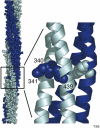
The glycine hinge of the kinase control module. (a) Atomic model of the kinase control module [25] illustrating the supercoiling of the two subunits (light and dark blue) and the location of the glycine hinge (box) that allows the four-helix bundle to bend ~10° [46]. (b) An expanded view of the hinge shows the helical backbones and highlights the six glycines of the hinge, residues G340, G341 and G439 in each subunit.

Similar articles

Cited by

References

    1. Bourret RB, Stock AM. Molecular information processing: lessons from bacterial chemotaxis. J. Biol. Chem. 2002;277:9625–9628. - PubMed
    1. Sourjik V. Receptor clustering and signal processing in E. coli chemotaxis. Trends Microbiol. 2004;12:569–576. - PubMed
    1. Parkinson JS, et al. Collaborative signaling by bacterial chemoreceptors. Curr. Opin. Microbiol. 2005;8:116–121. - PubMed
    1. Baker MD, et al. Systems biology of bacterial chemotaxis. Curr. Opin. Microbiol. 2006;9:187–192. - PubMed
    1. Szurmant H, Ordal GW. Diversity in chemotaxis mechanisms among the bacteria and archaea. Microbiol. Mol. Biol. Rev. 2004;68:301–319. - PMC - PubMed

Publication types

MeSH terms

LinkOut - more resources