

Nanoarchaeum equitans and Ignicoccus hospitalis: new insights into a unique, intimate association of two archaea
- PMID: 18165302
- PMCID: PMC2258681
- DOI: 10.1128/JB.01731-07
Nanoarchaeum equitans and Ignicoccus hospitalis: new insights into a unique, intimate association of two archaea
Abstract
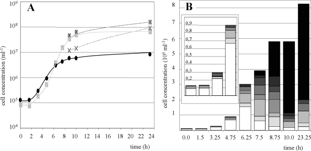
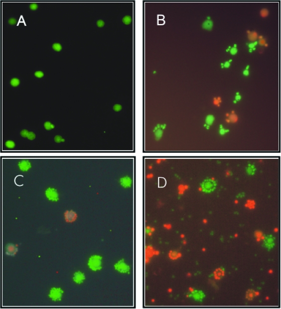
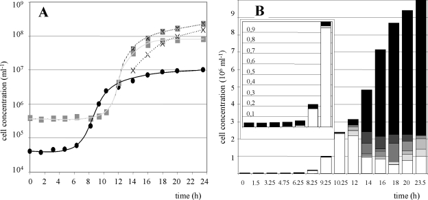
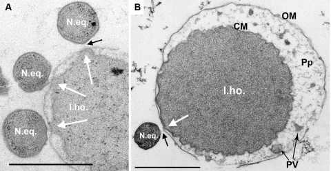
Nanoarchaeum equitans and Ignicoccus hospitalis represent a unique, intimate association of two archaea. Both form a stable coculture which is mandatory for N. equitans but not for the host I. hospitalis. Here, we investigated interactions and mutual influence between these microorganisms. Fermentation studies revealed that during exponential growth only about 25% of I. hospitalis cells are occupied by N. equitans cells (one to three cells). The latter strongly proliferate in the stationary phase of I. hospitalis, until 80 to 90% of the I. hospitalis cells carry around 10 N. equitans cells. Furthermore, the expulsion of H2S, the major metabolic end product of I. hospitalis, by strong gas stripping yields huge amounts of free N. equitans cells. N. equitans had no influence on the doubling times, final cell concentrations, and growth temperature, pH, or salt concentration ranges or optima of I. hospitalis. However, isolation studies using optical tweezers revealed that infection with N. equitans inhibited the proliferation of individual I. hospitalis cells. This inhibition might be caused by deprivation of the host of cell components like amino acids, as demonstrated by 13C-labeling studies. The strong dependence of N. equitans on I. hospitalis was affirmed by live-dead staining and electron microscopic analyses, which indicated a tight physiological and structural connection between the two microorganisms. No alternative hosts, including other Ignicoccus species, were accepted by N. equitans. In summary, the data show a highly specialized association of N. equitans and I. hospitalis which so far cannot be assigned to a classical symbiosis, commensalism, or parasitism.
Figures
References
-
- Baer, M. L., J. Ravel, S. A. Piñeiro, D. Guether-Borg, and H. N. Williams. 2004. Reclassification of salt-water Bdellovibrio sp. as Bacteriovorax marinus sp. nov. and Bacteriovorax litoralis sp. nov. Int. J. Syst. Evol. Microbiol. 541011-1016. - PubMed
-
- Baumann, P., and N. A. Moran. 1997. Non-cultivable microorganisms from symbiotic associations of insects and other hosts. Antonie van Leeuwenhoek 7239-48. - PubMed
-
- Burghardt, T., D. J. Näther, B. Junglas, H. Huber, and R. Rachel. 2007. The dominating outer membrane protein of the hyperthermophilic Archaeum Ignicoccus hospitalis: a novel pore-forming complex. Mol. Microbiol. 63166-176. - PubMed
-
- Ciccarelli, F. D., T. Doerks, C. von Mering, C. J. Creevey, B. Snel, and P. Bork. 2006. Toward automatic reconstruction of a highly resolved tree of life. Science 3111283-1287. - PubMed
Publication types
MeSH terms
Substances
LinkOut - more resources
Full Text Sources
