. 2008 Mar;190(5):1595-604.
doi: 10.1128/JB.01718-07. Epub 2007 Dec 28.

The type IV secretion system component VirB5 binds to the trans-zeatin biosynthetic enzyme Tzs and enables its translocation to the cell surface of Agrobacterium tumefaciens

Affiliations

The type IV secretion system component VirB5 binds to the trans-zeatin biosynthetic enzyme Tzs and enables its translocation to the cell surface of Agrobacterium tumefaciens

Khaled Ahmed Aly et al. J Bacteriol. 2008 Mar.

Abstract

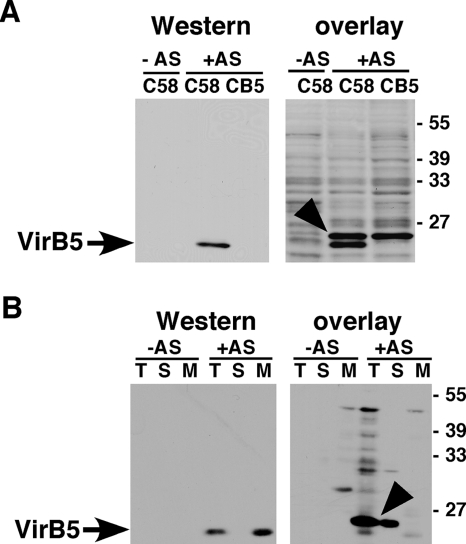
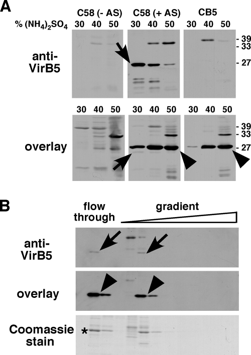
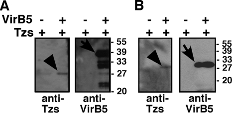
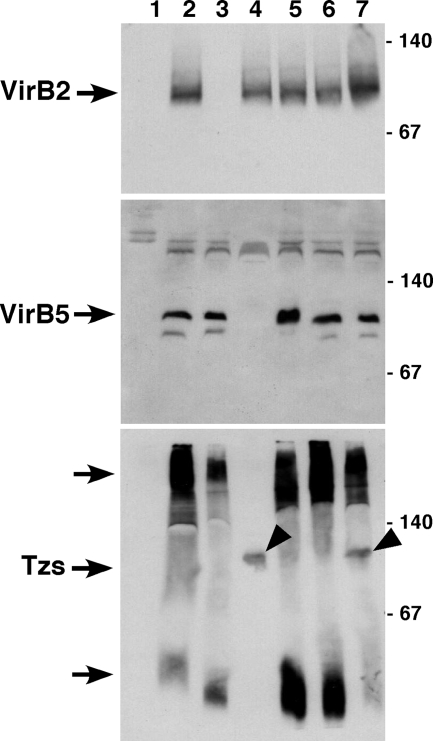
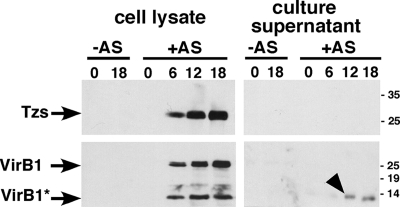
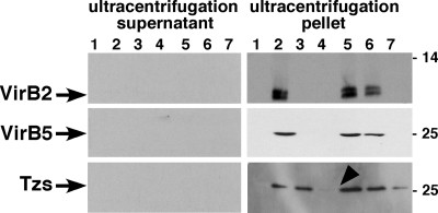
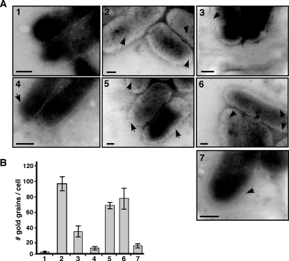
VirB5 is a minor component of the extracellular T pilus determined by the Agrobacterium tumefaciens type IV secretion system. To identify proteins that interact with VirB5 during the pilus assembly process, we purified VirB5 as a recombinant fusion protein and, by using a gel overlay assay, we detected a 26-kDa interacting protein in Agrobacterium cell lysates. The VirB5-binding protein was purified from A. tumefaciens and identified as the cytokinin biosynthetic enzyme Tzs. The VirB5-Tzs interaction was confirmed using pulldown assays with purified proteins and the yeast two-hybrid system. An analysis of the subcellular localization in A. tumefaciens showed that Tzs was present in the soluble as well as the membrane fraction. Tzs was extracted from the membranes with the mild detergent dodecyl-beta-D-maltoside in complexes of different molecular masses, and this association was strongly reduced in the absence of VirB5. Using immunoelectron microscopy, we also detected Tzs on the Agrobacterium cell surface. A functional type IV secretion system was required for efficient translocation to the surface, but Tzs was not secreted into the cell supernatant. The fact that Tzs localizes on the cell surface suggests that it may contribute to the interaction of Agrobacterium with plants.


Figures

FIG. 1.
FIG. 1.
Detection of a VirB5-binding protein using overlay assays. Wild-type C58 and the virB5 deletion mutant CB1005 carrying pPZP300 were grown under virulence gene-inducing (+AS) or noninducing conditions (−AS). For subcellular fractionation, the cells were lysed in a French press, followed by separation of the total cell lysate (T) into soluble (S) and membrane fractions (M). (A) Cell lysates were separated by SDS-PAGE, followed by Western blotting with VirB5-specific antiserum (left panel). The overlay assay was conducted by incubating the PVDF membrane after electrotransfer of the proteins with purified His6TrxAVirB5, followed by washing and Western blotting with VirB5-specific antiserum (right panel). (B) To analyze the subcellular localization of VirB5 and the interacting protein, samples of the T, S, and M fractions were separated by SDS-PAGE, followed by Western blotting (left panel) or the overlay assay was conducted, followed by Western blotting as described above (right panel). Arrows indicate VirB5, and arrowheads indicate the His6TrxAVirB5-binding protein detected in the overlay assay; numbers on the right indicate the molecular masses of reference proteins.
FIG. 2.
FIG. 2.
Purification of the VirB5-binding protein from the soluble fraction of C58 cell lysates. Wild-type C58 and the virB5 deletion variant CB1005 were grown under virulence gene-inducing (+AS) or noninducing conditions (−AS). (A) Cells were lysed in a French press and the soluble fractions were separated from the membranes by ultracentrifugation, followed by precipitation of proteins with increasing concentrations of (NH4)2SO4 as indicated. The precipitates were analyzed by SDS-PAGE and Western blotting with VirB5-specific antiserum (upper panel) or by overlay assay with His6TrxAVirB5, followed by the detection of bound VirB5 (lower panel). (B) Proteins precipitated with 40% (NH4)2SO4 from C58 extracts were dialyzed and applied to a MonoQ anion exchange column, and the fractions eluted from the column (flowthrough and NaCl gradient) were analyzed by SDS-PAGE and Western blotting, overlay assay and detection of VirB5, or Coomassie staining. The protein indicated by the asterisk was electrotransferred to a PVDF membrane and subjected to Edman sequencing. Arrows show VirB5, and arrowheads indicate the His6TrxAVirB5-binding protein detected using the overlay assay; numbers on the right indicate the molecular masses of reference proteins.
FIG. 3.
FIG. 3.
Analysis of the VirB5-Tzs interaction by pulldown assays. Purified Tzs was incubated with affinity bead-bound StrepIIVirB5 and His6TrxAVirB5 fusion proteins or with affinity beads alone, followed by sedimentation of the beads, washing, elution, and analysis of the bead-bound material by SDS-PAGE and Western blotting with specific antisera. (A) Analysis of proteins bound to Ni-nitrilotriacetic acid Sepharose after elution with imidazole. (B) Analysis of proteins bound to StrepTactin magnetic beads after elution with biotin. Arrows indicate VirB5 fusion proteins, and arrowheads indicate Tzs eluted from the affinity matrices. Molecular masses of reference proteins are shown on the right. −, absence of; +, presence of.
FIG. 4.
FIG. 4.
Analysis of DDM-extracted membrane protein complexes by blue native electrophoresis. Strains C58, CB1002 (ΔvirB2), CB1005 (ΔvirB5), and CB1008 (ΔvirB8) and complemented variants were cultivated on AB minimal medium in the absence (−AS, lane 1) or in the presence of AS (+AS, lanes 2 to 7) for virulence gene induction, followed by cell lysis, sedimentation of the membranes and extraction with 2% DDM. The samples were separated by blue native PAGE on a 15% gel, followed by Western blotting with VirB2-, VirB5-, and Tzs-specific antiserum. Lanes: 1, C58 without AS; 2, C58 with AS; 3, CB1002; 4, CB1005; 5, CB1002 pTrcB2; 6, CB1005 pTrcB5; 7, CB1008. Arrowheads point to Tzs in a 100-kDa complex in CB1005 and CB1008, and molecular masses of reference proteins are shown on the right (in kilodaltons). This experiment was conducted twice with qualitatively similar results.
FIG. 5.
FIG. 5.
Analysis of proteins secreted from A. tumefaciens. Strain C58 was cultivated in liquid AB minimal medium in the absence (−) or in the presence (+) of AS for virulence gene induction. The presence of VirB1, VirB1*, and Tzs was monitored in cell lysates as well as in culture supernatants at different time points after the induction of virulence gene expression (0 h, 6 h, 12 h, and 18 h). Samples were separated by SDS-PAGE, followed by Western blotting with VirB1 and Tzs-specific antiserum. The arrowhead points to secreted VirB1*, and molecular masses of reference proteins are shown on the right (in kilodaltons).
FIG. 6.
FIG. 6.
Analysis of the composition of T-pilus fractions. Strains C58, CB1002 (ΔvirB2), CB1005 (ΔvirB5), and CB1008 (ΔvirB8) and complemented variants were cultivated on AB minimal medium in the absence (−) of AS (lane 1) or in the presence (+) of AS (lanes 2 to 7) for virulence gene induction, followed by shearing of cells and ultracentrifugation for separation of extracellular high-molecular-mass structures (ultracentrifugation pellet) and low-molecular-mass proteins released from the cells (ultracentrifugation supernatant). Samples were separated by SDS-PAGE, followed by Western blotting with VirB2-, VirB5-, and Tzs-specific antiserum. Lanes: 1, C58 without AS; 2, C58 with AS; 3, CB1002; 4, CB1005; 5, CB1002 pTrcB2; 6, CB1005 pTrcB5; 7, CB1008. The arrowhead points to the reduced amount of Tzs in the ultracentrifugation pellet from CB1005, and molecular masses of reference proteins are shown on the right (in kilodaltons).
FIG. 7.
FIG. 7.
Immunoelectron microscopy detects Tzs on the A. tumefaciens cell surface. Strains C58, CB1002 (ΔvirB2), CB1005 (ΔvirB5), and CB1008 (ΔvirB8) and complemented variants were cultivated on AB minimal medium in the absence or in the presence of AS for virulence gene induction, followed by immunoelectron microscopy with Tzs-specific primary antibody and 10 nm gold-labeled secondary antibody. (A) Representative images of transmission electron micrographs; arrowheads point to gold grains on the cell surfaces of samples as follows: 1, C58 without AS; 2, C58 with AS; 3, CB1002; 4, CB1005; 5, CB1002 pTrcB2; 6, CB1005 pTrcB5; 7, CB1008. The contrast was increased to visualize the outline of cells for the purpose of presentation, but counting was conducted with reduced contrast settings that allowed the visualization of grains in even more heavily stained regions of the cells. Bars, 100 nm. (B) Quantification of results of the transmission electron microscopy analysis of Tzs on the cell surface; numbering of bars as for panel A. We counted 10 cells each from three independent induction experiments for each strain (total of 30 cells), and error bars show the standard deviations.

References

    1. Akiyoshi, D. E., H. Klee, R. M. Amasino, E. W. Nester, and M. P. Gordon. 1984. T-DNA of Agrobacterium tumefaciens encodes an enzyme of cytokinin biosynthesis. Proc. Natl. Acad. Sci. USA 815994-5998. - PMC - PubMed
    1. Akiyoshi, D. E., R. O. Morris, R. Hinz, B. S. Mischke, T. Kosuge, D. J. Garfinkel, M. P. Gordon, and E. W. Nester. 1983. Cytokinin/auxin balance in crown gall tumors is regulated by specific loci in the T-DNA. Proc. Natl. Acad. Sci. USA 80407-411. - PMC - PubMed
    1. Aly, K. A., and C. Baron. 2007. The VirB5 protein localizes to the T-pilus tips in Agrobacterium tumefaciens. Microbiology 1533766-3775. - PubMed
    1. Aragon, V., R. Diaz, E. Moreno, and I. Moriyon. 1996. Characterization of Brucella abortus and Brucella melitensis native haptens as outer membrane O-type polysaccharides independent from the smooth lipopolysaccharide. J. Bacteriol. 1781070-1079. - PMC - PubMed
    1. Atmakuri, K., E. Cascales, and P. J. Christie. 2004. Energetic components VirD4, VirB11 and VirB4 mediate early DNA transfer reactions required for bacterial type IV secretion. Mol. Microbiol. 541199-1211. - PMC - PubMed

Publication types

MeSH terms

LinkOut - more resources