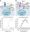
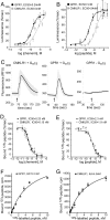
. 2008 Jan 8;105(1):64-9.
doi: 10.1073/pnas.0710487105. Epub 2007 Dec 28.

The genetic design of signaling cascades to record receptor activation

Affiliations

The genetic design of signaling cascades to record receptor activation

Gilad Barnea et al. Proc Natl Acad Sci U S A. .

Abstract

We have developed an experimental strategy to monitor protein interactions in a cell with a high degree of selectivity and sensitivity. A transcription factor is tethered to a membrane-bound receptor with a linker that contains a cleavage site for a specific protease. Activation of the receptor recruits a signaling protein fused to the protease that then cleaves and releases the transcription factor to activate reporter genes in the nucleus. This strategy converts a transient interaction into a stable and amplifiable reporter gene signal to record the activation of a receptor without interference from endogenous signaling pathways. We have developed this assay for three classes of receptors: G protein-coupled receptors, receptor tyrosine kinases, and steroid hormone receptors. Finally, we use the assay to identify a ligand for the orphan receptor GPR1, suggesting a role for this receptor in the regulation of inflammation.


Conflict of interest statement

The authors declare no conflict of interest.

Figures
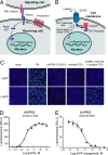
Fig. 1.
Fig. 1.
Design of the Tango assay. (A) A schematic of the Notch signaling pathway. The binding of Delta/Serrate/Lag-2 (DSL) ligands to Notch leads to a proteolytic cleavage of Notch by a protein complex involving presenilin. This cleavage releases a Notch intracellular domain (ICD) that translocates to the nucleus and modulates expression of target genes. (B) Schematic of the Tango assay method to monitor GPCR–arrestin interactions. Ligand binding to the target receptor stimulates recruitment of the arrestin-TEV protease fusion, triggering the release of the tethered transcription factor tTA. Free tTA then enters the nucleus and stimulates reporter gene activity. (C) Tango assay for the arginine vasopressin receptor AVPR2. HTL cells (an HEK293T-derived cell line containing a stably integrated tTA-dependent luciferase reporter) were transiently transfected with expression plasmids encoding the arginine vasopressin receptor 2 (AVPR2)-TEV cleavage site-tTA (AVPR2-TCS-tTA) and β-arrestin2-TEV protease (Arrestin-TEV) fusion proteins, as indicated. Control cells were transfected either with a tTA expression plasmid (tTA) or with empty parental expression plasmid (mock). Luciferase expression was detected by immunostaining with anti-luciferase (green), and cell nuclei were visualized with the DNA dye TOTO-3 (blue). Western blot analysis confirmed that receptor and arrestin components were expressed at similar levels in each transfection (data not shown). (D and E) Dose-response curves generated with a Tango assay for the human AVPR2. HTLA cells (an HEK293-derived cell line containing stable integrations of a tTA-dependent luciferase reporter and a β-arrestin2-TEV fusion gene) were transiently transfected with an AVPR2-TCS-tTA fusion gene. (D) Response to varying doses of the AVPR2 agonist vasopressin. (E) Response to varying doses of an AVPR2 antagonist, added 15 min before treatment with an EC80 dose (17 nM) of vasopressin. All errors bars represent SD (n = four measurements).
Fig. 2.
Fig. 2.
Tango assays for tyrosine kinase and steroid hormone receptors. (A) Receptor tyrosine kinase signaling. HTL cells were transfected with an IGF-1 receptor (IGF1R)–TCS-tTA fusion construct and a Shc1 PTB domain-TEV protease fusion plasmid. Luciferase activity in this IGF1R–Shc1 interaction assay was stimulated by IGF-1. (B) Ligand-induced homo- and heterodimerization of estrogen receptors (ERs). HTL cells were transfected with a CD8-ERα-TCS-tTA fusion and either an ERα- or ERβ-TEV protease fusion to measure ERα homodimerization (light circles) or ERα/ERβ heterodimerization (dark squares) as illustrated. Responses were normalized to the maximal response for each receptor. All error bars represent SD (n = four measurements).
Fig. 3.
Fig. 3.
Using the Tango assay to profile adrenergic receptor agonists and antagonists. (A and B) Agonist and antagonist selectivity profiling using Tango assays for human adrenergic receptors. HTLA cells were transiently transfected with α2A-, α2B-, α2C-, β1-, or β2-adrenergic receptor-TCS-tTA fusions, each of which contained the C-terminal tail sequence from AVPR2. (A) In these representative Tango agonist assays, α-adrenergic receptors were preferentially activated by the α-selective agonist UK14,304, and β-adrenergic receptors were preferentially activated by the β-selective agonist isoproterenol. (B) In representative antagonist assays, α- and β-ARs displayed the expected selectivity to the antagonists yohimbine and alprenolol, respectively. All error bars represent SD (n = four measurements). See SI Table 2 for complete results. (C) Stimulation of luciferase reporter gene activity in the β2-adrenergic receptor Tango assay by a full agonist, isoproterenol, and partial agonists salbutamol, clenbuterol, and nylidrin. Agonist-stimulated luciferase activity was measured in an HEK293T-derived cell line containing stably integrated luciferase reporter, β-arrestin2-TEV, and β2-AR-TCS-tTA (with AVPR2 C-terminal modification) constructs.
Fig. 4.
Fig. 4.
Identification of an agonist for the orphan receptor GPR1. (A and B) Dose-response profiles of GPR1 and CMKLR1 Tango assays in response to recombinant human chemerin protein (A) and a peptide fragment of chemerin (chemerin 145–157) (B). Responses were normalized to the maximal response for each receptor. (C) Calcium mobilization assay showing activation of CMKLR1 and GPR1 by chemerin peptide (chemerin 149–157, at 1 μM) in the presence of the promiscuous G protein Gα15. Time of ligand addition is indicated by the arrows. The responses of seven representative cells were averaged. All error bars represent SD. (D–G) Binding of radiolabeled chemerin C-terminal peptide to GPR1-and CMKLR1-transfected cells. Shown is displacement of iodinated chemerin C-terminal peptide (chemerin149–157) binding to GPR1- (black circles) and CMKLR1-expressing cells (gray squares) by full-length chemerin peptide (D) or unlabeled chemerin149–157 (E). Shown is saturation binding of 125I-chemerin149–157 to GPR1-transfected cells (F) and CMKLR1-transfected cells (G).

References

    1. Wehr MC, Laage R, Bolz U, Fischer TM, Grunewald S, et al. Monitoring regulated protein-protein interactions using split TEV. Nat Methods. 2006;3:985–993. - PubMed
    1. Struhl G, Adachi A. Nuclear access and action of notch in vivo. Cell. 1998;93:649–660. - PubMed
    1. Schroeter EH, Kisslinger JA, Kopan R. Notch-1 signalling requires ligand-induced proteolytic release of intracellular domain. Nature. 1998;393:382–386. - PubMed
    1. Gossen M, Bujard H. Tight control of gene expression in mammalian cells by tetracycline-responsive promoters. Proc Natl Acad Sci USA. 1992;89:5547–5551. - PMC - PubMed
    1. Parks TD, Leuther KK, Howard ED, Johnston SA, Dougherty WG. Release of proteins and peptides from fusion proteins using a recombinant plant virus proteinase. Anal Biochem. 1994;216:413–417. - PubMed

Publication types