. 2008 Jan 23;130(3):818-20.
doi: 10.1021/ja077082c. Epub 2008 Jan 1.

A single-molecule nanopore device detects DNA polymerase activity with single-nucleotide resolution

Affiliations

A single-molecule nanopore device detects DNA polymerase activity with single-nucleotide resolution

Scott L Cockroft et al. J Am Chem Soc. .

Abstract

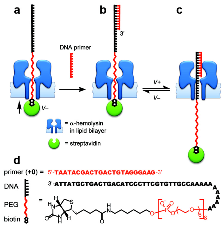
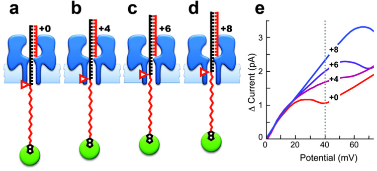
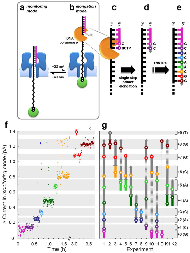
The ability to monitor DNA polymerase activity with single-nucleotide resolution has been the cornerstone of a number of advanced single-molecule DNA sequencing concepts. Toward this goal, we report the first observation of the base-by-base DNA polymerase activity with single-base resolution at the single-molecule level. We describe the design and characterization of a supramolecular nanopore device capable of detecting up to nine consecutive DNA polymerase-catalyzed single-nucleotide primer extensions with high sensitivity and spatial resolution (<or=2.4 A). The device is assembled in a suspended lipid membrane by threading and mechanically capturing a single strand of DNA-PEG copolymer inside an alpha-hemolysin protein pore. Single-nucleotide primer extensions result in successive displacements of the template DNA strand within the protein pore, which can be monitored by the corresponding stepped changes in the ion current flowing through the pore under an applied transmembrane potential. The system described thus represents a promising advance toward nanopore-mediated single-molecule DNA sequencing concept and, in addition, might be applicable to studying a number of other biopolymer-protein interactions and dynamics.


Figures

Figure 1
Figure 1
Fabrication and configuration of the interlocked α-HL•DNA-PEG transmembrane complex. (a) A single streptavidin-capped DNA-PEG strand on one side of the membrane is driven into, and held within an α-HL pore under an applied negative transmembrane potential. (b) Hybridization of a DNA primer to the protruding 5’-ssDNA region of the threading strand on the opposite side of the membrane, furnishes the fully interlocked α-HL•DNA-PEG complex. (c) The position of the threading strand held inside the pore can be flipped back and forth by changing the sign of the applied potential. (d) The composition of the threading DNA-PEG strand.
Figure 2
Figure 2
Establishing the device response to changes in primer length. The interlocked α-HL•DNA-PEG transmembrane complexes containing primers of different lengths. (a) 23 nucleotides (+0), (b) 27 nucleotides (+4), (c) 29 nucleotides (+6), and (d) 31 nucleotides (+8). All primer sequences are fully complementary to the 3’-end of the template sequence (Fig. 1). As the length of the primer increases, the ratio of PEG to DNA in the pore is increased (red pointers mark the PEG-DNA transition in each complex). (e) Primer length differences manifest themselves in the characteristic current-voltage (I–V) trace of each complex. The largest and most reproducible current difference between the four complexes was observed at +40 mV (vertical dotted line). Currents were averaged from the final 0.2 sec of the 2 sec ion current recording at each potential. The I–V traces were averaged from 10 or more recordings. Experiments were performed under conditions appropriate for DNA polymerase activity (150 mM KCl, 25 mM Tris, 4.5 mM MgCl2, at pH 8.0 and 22 ± 2 °C, see Fig. S1.
Figure 3
Figure 3
Monitoring DNA polymerase-catalyzed single-nucleotide primer extensions. (a) The length of the DNA primer in a fully interlocked α-HL•DNA-PEG complex is reported by ion current measurements in the monitoring mode (+40 mV). (b) In the elongation mode (−30 mV), the 3’-OH of the primer is accessible to the DNA polymerase. (c) DNA polymerase bound to the starting primer-template complex, with the correct incoming deoxynucleotide triphosphate. (d) DNA polymerase catalyzes the elongation of the primer by incorporating a single dNTP against the DNA template threaded through the pore. (e) Templated extension of the primer sequence after multiple base incorporation steps. (f) A plot of cumulative change in the ion channel current recorded in the monitoring mode versus time. The data contains nine distinct current levels corresponding to the starting primer (+0, Fig. 2A), the seven intermediate polymerized states, and the extended primer (+8, Fig. 2d). Primer extension was paused for over one hour after the incorporation of 6 bases (+6) until the required dGTP was added (note the broken time axis). (g) Alignment of multiple experiments initiated with primers of different lengths (vertical colored bars). The dark gray arrows indicate the direction of primer extension. The white dots in colored circles represent the average cumulative ion current for each length of primer (s.e.m. ±0.05 pA). The horizontal light gray stripes indicate the average current maxima and minima for each primer length. The numbers to the right indicate the base extension relative to the shortest primer (+0), and the letters in parentheses identify the base at the 3’ end of each primer. Dashed lines represent nucleotide incorporation steps that were not sampled by current recordings. Experiment 1 corresponds directly to the current recordings shown in Fig. 3F. Experiment D shows the displacement of a short primer (+0) by a longer primer (+8) as described in the main text. TopoTaq DNA polymerase was used in experiments 1 to 11. The Klenow fragment (exo-) of E. coli DNA polymerase I was used in experiments K1 and K2. Polymerization was terminated with the appropriate dideoxynucleotide triphosphate (ddNTP) in experiments 2, 7, and K2. Experimental conditions were as described in Fig. 2, but with the addition of 2 nM DNA polymerase and 0.01–0.2 mM dNTPs. See Fig. S4–S17 for complete current versus time recordings for all experiments.

Similar articles

Cited by

References

    1. Shendure J, Mitra RD, Varma C, Church GM. Nat. Rev. Genet. 2004;5:335–344. - PubMed
    2. Bayley H. Curr. Opin. Chem. Biol. 2006;10:628–637. - PubMed
    1. Maier B, Bensimon D, Croquette V. Proc. Natl. Acad. Sci. USA. 2000;97:12002–12007. - PMC - PubMed
    2. Braslavsky I, Hebert B, Kartalov E, Quake SR. Proc. Natl. Acad. Sci. USA. 2003;100:3960–3964. - PMC - PubMed
    3. Bustamante C, Bryant Z, Smith SB. Nature. 2003;421:423–427. - PubMed
    4. Abbondanzieri EA, Greenleaf WJ, Shaevitz JW, Landick R, Block SM. Nature. 2005;438:460–465. - PMC - PubMed
    5. Greenleaf WJ, Block SM. Science. 2006;313:801. - PMC - PubMed
    1. Sanchez-Quesada J, Saghatelian A, Cheley S, Bayley H, Ghadiri MR. Angew. Chem. Int. Ed. 2004;43:3063–3067. - PMC - PubMed
    2. Mathe J, Visram H, Viasnoff V, Rabin Y, Meller A. Biophys. J. 2004;87:3205–3212. - PMC - PubMed
    3. Nakane J, Wiggin M, Marziali A. Biophys. J. 2004;87:615–621. - PMC - PubMed
    4. Ashkenasy N, Sanchez-Quesada J, Bayley H, Ghadiri MR. Angew. Chem. Int. Ed. 2005;44:1401–1404. - PMC - PubMed
    1. Kasianowicz JJ, Brandin E, Branton D, Deamer DW. Proc. Natl. Acad. Sci. USA. 1996;93:13770–13773. - PMC - PubMed
    2. Vercoutere W, Winters-Hilt S, Olsen H, Deamer DW, Haussler D, Akeson M. Nat. Biotechnol. 2001;19:248–252. - PubMed
    3. Rhee M, Burns MA. Trends Biotechnol. 2006;24:580–586. - PubMed
    4. Astier Y, Braha O, Bayley H. J. Am. Chem. Soc. 2006;128:1705–1710. - PubMed
    1. Movileanu L, Howorka S, Braha O, Bayley H. Nat. Biotechnol. 2000;18:1091–1095. - PubMed
    2. Howorka S, Cheley S, Bayley H. Nat. Biotechnol. 2001;19:636–639. - PubMed
    3. Cheley S, Xie H, Bayley H. ChemBioChem. 2006;7:1923–1927. - PubMed
    4. Hornblower B, Coombs A, Whitaker RD, Kolomeisky A, Picone SJ, Meller A, Akeson M. Nat. Methods. 2007;4:315–317. - PubMed

Publication types

MeSH terms