. 2007 Dec 28;3(12):e227.
doi: 10.1371/journal.pgen.0030227.

Argonaute2 is essential for mammalian gastrulation and proper mesoderm formation

Affiliations

Argonaute2 is essential for mammalian gastrulation and proper mesoderm formation

Reid S Alisch et al. PLoS Genet. .

Abstract

Mammalian Argonaute proteins (EIF2C1-4) play an essential role in RNA-induced silencing. Here, we show that the loss of eIF2C2 (Argonaute2 or Ago2) results in gastrulation arrest, ectopic expression of Brachyury (T), and mesoderm expansion. We identify a genetic interaction between Ago2 and T, as Ago2 haploinsufficiency partially rescues the classic T/+ short-tail phenotype. Finally, we demonstrate that the ectopic T expression and concomitant mesoderm expansion result from disrupted fibroblast growth factor signaling, likely due to aberrant expression of Eomesodermin. Together, these data indicate that a factor best known as a key component of the RNA-induced silencing complex is required for proper fibroblast growth factor signaling during gastrulation, suggesting a possible micro-RNA function in the formation of a mammalian germ layer.


Conflict of interest statement

Competing interests. The authors have declared that no competing interests exist.

Figures

Figure 1
Figure 1. Characterization of the Ago2 Disruption in ES Cell Clone RRE192
(A) Insertion of the gene-trap vector into intron 12 of the mouse Ago2 locus. Exons 1, 12, and 18 are labeled. Exons encoding the PAZ domain are shown in yellow, and exons encoding the PIWI domain are shown in orange. The insertion cassette contains a splice acceptor (SA), a fusion of the β-galactosidase and neomycin phosphotransferase coding sequences (β-geo), and a polyadenylation signal (pA). FRT and loxP sites are denoted as black triangles. The relative location of primers used for genotyping are shown as half-arrows and are labeled a, b, and c. (B) The genotypes of embryos from heterozygous intercrosses. Shown is a gel displaying PCR products using primers a and c, identifying the normal allele, and primers a and b, identifying the interrupted allele. The PCR loaded into the water lane lacked template DNA and acts as a negative control. +/+ represents wild-type; +/– represents Ago2 heterozygote; –/– represents Ago2 homozygous mutant. (C) Western blot analysis of AGO2 in e7.5 wild-type (+/+) and Ago2 homozygous mutant (–/–) embryos. EIF4E was used as a loading control. (D) Variable phenotype of Ago2 homozygous (Ago2–/–) mutant embryos at e9.5. Shown here are three littermates. The variable Ago2–/– phenotypes included deciduas containing only embryonic remnants (ii and iii). This phenotype also was variable as either remnants of embryoid structures (ii) or as cell masses lacking obvious embryonic development (iii). Note the size magnification of the embryonic remnants, as they are much smaller than the wild-type (WT) littermate (i).
Figure 2
Figure 2. The Homozygous Disruption of Ago2 Results in an Expansion of T Expression and Mesoderm Formation
(A, B) Whole-mount in situ hybridization using an antisense probe against T on e7.5 wild-type (A) and Ago2 –/– (B) embryo littermates. The Ago2 –/– embryos exhibit an expansion of the primitive streak (block-arrow). Note that Ago2 –/– e7.5 embryos are smaller and rounder than wild-type, suggesting aberrant growth. The scale bar represents 200 μm. (A, B, insets) Sections from whole-mount in situ hybridized e7.5 embryos. Shown are representative wild-type (A, inset) and Ago2 –/– embryos (B, inset). The scale bar represents 50 μm. (C−H) Paraffin sections from Ago2+/ (C, E, G) and Ago2 –/– (D, F, H) e7.5 embryos were stained with antibodies against β-galactosidase (C, D, G, H; green) and BRACHYURY (E, F, G, H; red). Coexpression of the proteins will appear yellow (G, H; merge). At this stage, wild-type Ago2 expression is restricted to the epithelial cell layer, and it does not overlap with BRACHYURY in the primitive streak. The scale bar represents 50 μm. The arrows denote the relative location of the primitive streak. The brackets indicate the approximate region of the mesoderm layer and/or the epithelial cell layer. m = mesoderm layer; ec = epithelial cell layer.
Figure 3
Figure 3. The Distribution of Tail Length for Each Genotype
(A) Shown are four mice from the same litter. While the tail lengths are indistinguishable between wild-type (WT) and Ago2 heterozygote (Ago2+/ ) mice, the T heterozygote (T/+) tail is reduced to approximately 30% of wild-type. In contrast, double heterozygous (Ago2+/ T/+) mice have tail lengths that are approximately 60% of wild-type. (B) Shown are the raw data (vertical scatterplot) overlaid with a notched-box plot. The center of the notched-box plot is the median, and the endpoints of the notches are located at the median confidence intervals. The extreme endpoints of the notched-box plot represent the 25% (lower) and the 75% (upper) quartiles of the scatter plot data. The x-axis shows each genotype name, and n is the number of mice. The y-axis shows the ratio of tail-to-body length. The asterisks denote that the double heterozygotes had significantly greater tail-to-body length ratios relative to single T heterozygotes (p = 0.007).
Figure 4
Figure 4. The Homozygous Disruption of Ago2 Results in a Disruption of FGF Signaling
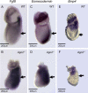
(A−F) Whole-mount in situ hybridization using an antisense probe against Fgf8, Eomesodermin, or Bmp4 on e7.5 wild-type (A, C, E) and Ago2 –/– (B, D, F) embryo littermates. The Ago2 –/– embryos exhibit a lateral expansion of Fgf8 and Eomesodermin expression away from the primitive streak (B, D; block-arrow). In contrast, the localization of Bmp4 expression is indistinguishable between wild-type (E) and Ago2 –/– (F) embryo littermates. (A−D) Embryos imaged with reflective light. (E, F) Embryos imaged with reflective and transmitted light. The scale bar represents 200 μm.
Figure 5
Figure 5. A Working Model for Brachyury (T) Induction at the Commencement of Mouse Gastrulation
In wild-type mice, Ago2 regulates the proper level of Eomesodermin (Eomes) gene expression, which ultimately induces the downstream expression of Fgf8 and T. In the absence of Ago2, Eomes is not properly regulated and becomes overexpressed, resulting in the downstream overinduction of Fgf8 and T. Other possible models are described in the text.

References

    1. Parker JS, Roe SM, Barford D. Crystal structure of a PIWI protein suggests mechanisms for siRNA recognition and slicer activity. EMBO J. 2004;23:4727–4737. - PMC - PubMed
    1. Song JJ, Smith SK, Hannon GJ, Joshua-Tor L. Crystal structure of Argonaute and its implications for RISC slicer activity. Science. 2004;305:1434–1437. - PubMed
    1. Lingel A, Simon B, Izaurralde E, Sattler M. Structure and nucleic-acid binding of the Drosophila Argonaute 2 PAZ domain. Nature. 2003;426:465–469. - PubMed
    1. Yan KS, Yan S, Farooq A, Han A, Zeng L, et al. Structure and conserved RNA binding of the PAZ domain [erratum appears in Nature. 2004 Jan 15;427(6971):265] Nature. 2003;426:468–474. - PubMed
    1. Song JJ, Liu J, Tolia NH, Schneiderman J, Smith SK, et al. The crystal structure of the Argonaute2 PAZ domain reveals an RNA binding motif in RNAi effector complexes. Nat Struct Biol. 2003;10:1026–1032. - PubMed

Publication types