. 2007 Dec 28;3(12):e235.
doi: 10.1371/journal.pgen.0030235.

Deletion of the MBII-85 snoRNA gene cluster in mice results in postnatal growth retardation

Affiliations

Deletion of the MBII-85 snoRNA gene cluster in mice results in postnatal growth retardation

Boris V Skryabin et al. PLoS Genet. .

Abstract

Prader-Willi syndrome (PWS [MIM 176270]) is a neurogenetic disorder characterized by decreased fetal activity, muscular hypotonia, failure to thrive, short stature, obesity, mental retardation, and hypogonadotropic hypogonadism. It is caused by the loss of function of one or more imprinted, paternally expressed genes on the proximal long arm of chromosome 15. Several potential PWS mouse models involving the orthologous region on chromosome 7C exist. Based on the analysis of deletions in the mouse and gene expression in PWS patients with chromosomal translocations, a critical region (PWScr) for neonatal lethality, failure to thrive, and growth retardation was narrowed to the locus containing a cluster of neuronally expressed MBII-85 small nucleolar RNA (snoRNA) genes. Here, we report the deletion of PWScr. Mice carrying the maternally inherited allele (PWScr(m-/p+)) are indistinguishable from wild-type littermates. All those with the paternally inherited allele (PWScr(m+/p-)) consistently display postnatal growth retardation, with about 15% postnatal lethality in C57BL/6, but not FVB/N crosses. This is the first example in a multicellular organism of genetic deletion of a C/D box snoRNA gene resulting in a pronounced phenotype.


Conflict of interest statement

Competing interests. The authors have declared that no competing interests exist.

Figures
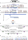
Figure 1
Figure 1. Genome Organization of the PWS/AS Locus in Man and Mouse
(A) Structure of human chromosome 15. (B) Schematic representation of the human 15q11-q13 region (not drawn to scale). The centromeric (cen) and telomeric (tel) positions are labeled. PWS protein coding genes and snoRNA genes are marked as boxes and ovals, respectively. Paternally, maternally, and biparentally expressed genes are labeled blue, red, and white, respectively. IPW exons of the paternally expressed, imprinted U-UBE3A-AS transcript are indicated by green bars and a blue arrow, respectively. The imprinting center (IC) is indicated with a small circle. The snoRNA genes: HBII-436, HBII-13, HBII-437, HBII-438A, HBII-85. HBII-52 and HBII-438B are labeled as 436, 13, 437, 438A, 85, 52 and 438B, accordingly. (C) Corresponding mouse syntenic chromosome 7C region. The paternally expressed Lncat transcript is indicated by a blue arrow. (D) Deletions in the PWS/AS region. (1) Insertion of the Epstein–Barr virus Latent Membrane Protein 2A, (LMP2A) transgene into the PWS/AS locus resulted in deletion of approximately 4 megabases between the Mlsn1 and Gas2 genes [12,29]. Depending on the parental origin it causes PWS or AS symptoms in mice. (2) The 42 kb Snrpn promoter PWS-IC deletion resulted in loss of imprinting and a PWS-like phenotype in mice [13]; analogous deletion of the PWS-IC and Snrpn gene up to exon 7 results in a paternal to maternal imprint switch and leads to PWS [40]. (3) Paternal Snrpn exon 2 deletion [14] produced no abnormal phenotype. (4) Mice with a deletion of 1kb (exons 5–7) of the Snrpn gene are phenotypically normal [13]. (5) Paternal deletion from Snrpn to Ube3a resulted in a PWS-like phenotype in mice [14]. (6) Deletion affecting npc Ipw exons and MBII-52 snoRNA gene cluster after paternal inheritance produced no PWS-like phenotype [15]. (7) Targeting of the Necdin gene produced either a PWS-like phenotype [21,23] or no obvious phenotype [22]. The discrepancies were explained by different strategies in Necdin targeting and different mouse genetic backgrounds [15]. (8) Inactivation of the Magel2 gene produced altered behavioral rhythmicity [19]. (9) Deletion of the Mkrn3 (Zfp127) gene had no apparent phenotypic effect [15]. (10) PWS critical region (PWScr). It has been proposed that deletion of this region should play a major role in the development of PWS [16,24]. Paternal deletion of PWScr causes growth retardation in mice (this study). (E) Locus containing the cluster of MBII-85 snoRNA genes and Ipw exons, and its deletion targeting strategy using “chromosome engineering” [26]. Filled boxes represent 5′HR and 3′HR DNA probes (labeled green and purple respectively). Targeting constructs 5′HPRT/PWScr_targ and 3′HPRT/PWScr_targ are indicated. Open boxes correspond to the Thymidine kinase gene (TK), Neomycin resistance gene (Neo), Puromycin resistance gene (Puro), as well as the 5′ and 3′ parts of the Hypoxanthine Phosphoribosyl Transferase gene (HPRT). LoxP sites are indicated by arrows. (F) Structure of the PWScr after two consecutive HR events, and CRE recombinase-induced deletion. (G) Sequencing of integration sites after HR and deletion of the PWScr confirmed the anticipated deletion at the nucleotide level. Extended sequences (Genbank accession number EU233428) of regions flanking the HPRT cassette insertion are presented in Figure S1.
Figure 2
Figure 2. Analysis of Mice with the PWScr Deletion
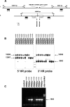
(A) Schematic representation of 1. wild-type and 2. PWScr targeted alleles. DNA fragments are labeled according to Figure 1E and 1F. EcoRV (RV) restriction endonuclease sites are designated by vertical arrows. Sizes of diagnostic restriction fragments are indicated. The positions of PCR primers MB85deld1 and MB85delr1 are shown. (B) Southern blot analysis. EcoRV digested DNA samples of individual mice (genotype indicated on top) were hybridized with 5′HR and 3′HR probes. Bands representing the wild-type alleles (12987 bp for 5′HR and 9063 bp for 3′HR) and the PWScr-deleted allele (19998 bp) are indicated by arrows. Size markers (14140, 8453, 7242, 6369, and 5687 bp) are represented by horizontal bars between the two blots. (C) Genotyping by PCR analysis using primers MB85deld1/MB85delr1 (positions are shown in A). Deletion of PWScr results in a PCR product of 1665 bp. Genotypes of mice are indicated.
Figure 3
Figure 3. Growth Differences among PWScrm+/p− and PWScrm+/p+ Siblings
Representative pair of mice from the same litter at postnatal day 10 (129SV x C57BL/6 genetic crosses).
Figure 4
Figure 4. Growth Retardation and Postnatal Lethality in PWScrm+/p− Mice (129SV x C57BL/6 Genetic Background)
(A) Growth dynamics of 82 investigated male mice. The yellow line corresponds to weight gain of 34 mice with the PWScr-deleted allele; black bars are statistically significant intervals (confidence level 95%, p=0.05). The black line corresponds to 48 wild-type males; black bars are statistically significant intervals (p=0.05). (B) Growth dynamics of 82 investigated female mice. The yellow line corresponds to weight gain in 38 females with the PWScr-deleted allele; black bars are statistically significant intervals (p=0.05). The black line corresponds to 44 wild-type females; black bars are statistically significant intervals (p=0.05). (C) Growth dynamics of 40 female mice with the maternally transmitted PWScr-deleted allele. The red line corresponds to weight gain in 16 females with the PWScr-deleted allele; green bars are statistically significant intervals (p=0.05). The black line corresponds to 24 wild-type females. (D) Growth comparison of 15-day-old control mice with wild-type mice. Mice, containing the 5′HPRT/PWScr_targ cassette in the PWScr locus (5′HPRTm−/p+; 6.898±0.257 g), are represented by white bars, and wild-type mice (6.711±0.129 g) by black bars (p=0.2751). (E) Embryo weights (E18.5) and the corresponding placenta weights (n=15 for all groups). The weights of embryos and placentas from PWScrm+/p+ and PWScrm+/p− mice are indicated with black and yellow bars, respectively. Embryo weights E18.5 PWScrm+/p+ 1.290±0.105 g (mean±SD), PWScrm+/p− 1.253±0.151 g, (p=0,934); placenta weights E18.5 PWScrm+/p+ 0.086±0.012 g, PWScrm+/p− 0.094±0.021 g, (p=0.290). (F) Postnatal lethality of PWScrm+/p− (n=64) and wild type PWScrm+/p+ (n=57) mice in 129SV x C57BL/6 (>85% C57BL/6 contribution) genetic crossings (p<0.02). White and black bars represent percentages of surviving and dead mice, respectively, after 90 days.
Figure 5
Figure 5. mRNA Analysis of PWS and AS Imprinted Genes in PWScr-Deleted Mice
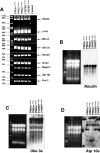
(A) RT-PCR analysis. Mouse genotypes are indicated. 5′HPRT+/+ and 3′HPRT+/+ correspond to homozygous 5′HPRT/PWScr_targ or 3′HPRT/PWScr_targ targeted mice, respectively; size markers are given in bp at the left; the position of RT-PCR products corresponding to individual transcripts are marked by arrows. Gapdh and β-Act transcripts were used as controls. (B–D) Northern blot analyses of selected mRNAs from the PWS locus. Ethidium bromide-stained RNA gels (prior to blotting) are shown as RNA loading controls. RNA size markers are as follows: 6000, 4000, 3000, 2000, 1500, 1000, 500, and 200 nt.
Figure 6
Figure 6. Expression of snoRNAs and Ipw Transcripts in PWScr-Deleted Mice
(A) Expression analysis of snoRNAs from the PWS locus. 5.8S rRNA was probed as RNA loading control. Mouse genotypes are indicated (see Figure 5). (B) Northern blot analysis of Ipw transcripts. Ethidium bromide-stained RNA gels (prior to blotting) are shown as RNA loading controls. RNA size markers are identical to those in Figure 5.
Figure 7
Figure 7. Expression of Ipw Exons and MBII-52 snoRNA throughout Different Stages of Wild-Type Mouse Development (Two Independent Experiments Using RNAs from WT Mice)
We examined the expression patterns of Ipw exons (Lncat derived ESTs) and MBII-52 snoRNAs by Northern blot hybridization using total RNA from embryonal (E) and postnatal (P) brain, except for embryonic days 10.5 and 12.5, when total RNA was isolated from whole embryos. As loading control 5.8S rRNA was probed. Signals appeared between E12.5 and E15.5 days (note that this observation may, in part, be due to the fact that we switched from whole embryos to brains as sources of total RNA). Interestingly, while expression of MBII-52 snoRNA constantly increased, the levels of transcripts harboring Ipw exons fluctuated. This might indicate that maturation of MBII-52 snoRNA is not dependent solely on Ipw exon processing and that snoRNAs might be derived from Lncat npcRNA via alternative pathways.

References

    1. Kantor B, Shemer R, Razin A. The Prader-Willi/Angelman imprinted domain and its control center. Cytogenet Genome Res. 2006;113:300–305. - PubMed
    1. Nicholls RD, Knepper JL. Genome organization, function, and imprinting in Prader-Willi and Angelman syndromes. Annu Rev Genomics Hum Genet. 2001;2:153–175. - PubMed
    1. Burd L, Vesely B, Martsolf J, Kerbeshian J. Prevalence study of Prader-Willi syndrome in North Dakota. Am J Med Genet. 1990;37:97–99. - PubMed
    1. Whittington JE, Holland AJ, Webb T, Butler J, Clarke D, et al. Population prevalence and estimated birth incidence and mortality rate for people with Prader-Willi syndrome in one UK Health Region. J Med Genet. 2001;38:792–798. - PMC - PubMed
    1. Wattendorf DJ, Muenke M. Prader-Willi syndrome. Am Fam Physician. 2005;72:827–830. - PubMed

Publication types

Substances