. 2008 Jan;198(1):122.e1-8.
doi: 10.1016/j.ajog.2007.06.065.

Experimental amniotic fluid infection in sheep: effects of Ureaplasma parvum serovars 3 and 6 on preterm or term fetal sheep

Affiliations

Experimental amniotic fluid infection in sheep: effects of Ureaplasma parvum serovars 3 and 6 on preterm or term fetal sheep

Timothy J M Moss et al. Am J Obstet Gynecol. 2008 Jan.

Abstract

Objective: The objective of the study was to determine the effects in late gestation of Ureaplasma parvum serovar 3 colonization and the effects, preterm, of U. parvum serovar 6.

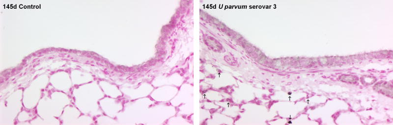
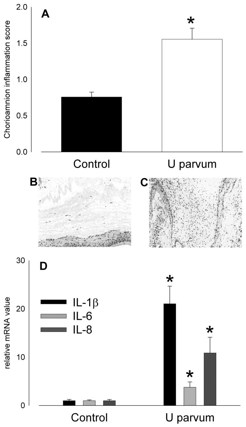
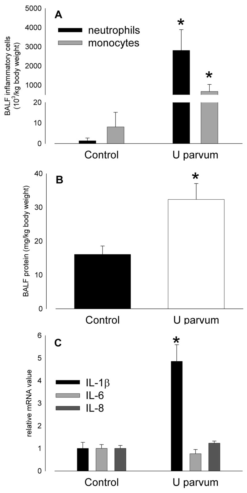
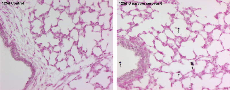
Study design: Ewes received an intraamniotic (i.a.) injection of U. parvum serovar 6 (20 x 10(6) colony-forming units [cfu]; n = 9), U. parvum serovar 3 (20 x 10(3) cfu; n = 6), vehicle (n = 10), or saline (n = 4) on day 80 of pregnancy (d). The lambs were delivered at 125 d (U. parvum serovar 6, n = 9; saline or media controls, n = 9) or 145 d (U. parvum serovar 3, n = 6; media controls, n = 5) for assessment of inflammation and lung maturation.

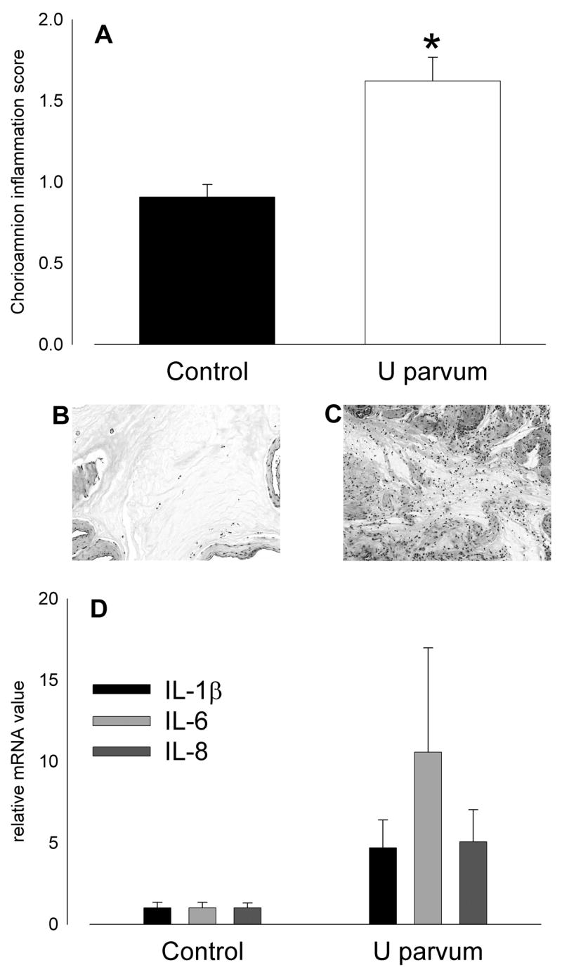
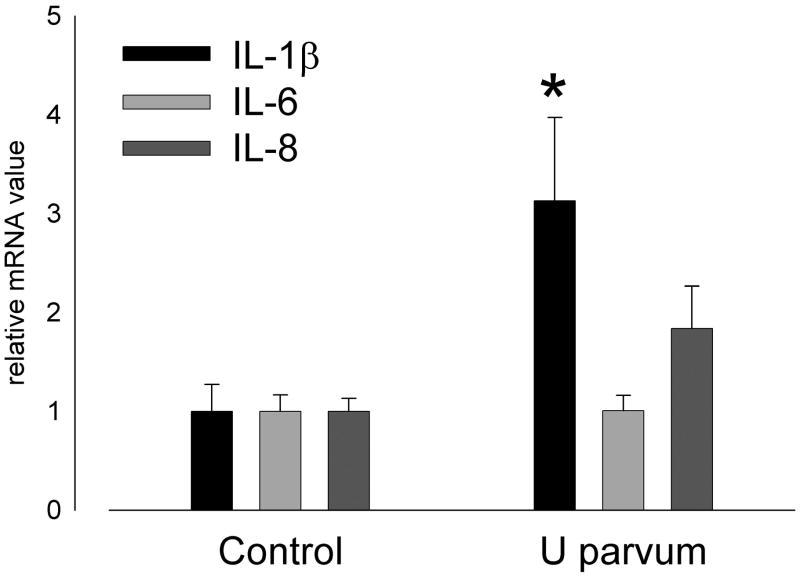
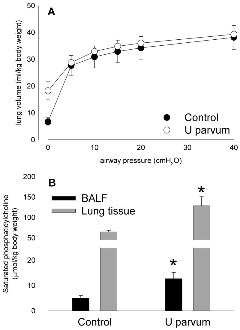
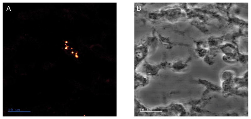
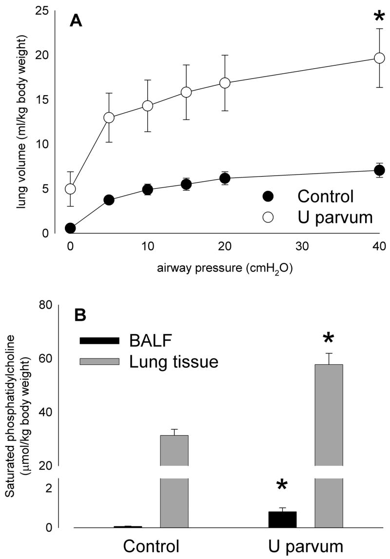
Results: I.a. ureaplasmas caused histologic chorioamnionitis but not preterm delivery. Fetal lung epithelium was colonized with ureaplasmas at both gestational ages, and pulmonary interleukin-8 levels had doubled in the ureaplasma-colonized animals, compared with the controls at 145 d. Surfactant levels in bronchoalveolar lavage fluid had increased 8-fold and 2.5-fold at 125 and 145 d, respectively, after ureaplasma injection.

Conclusion: Fetal lung inflammation and altered development accompanies ureaplasma colonization, regardless of age at delivery.


Figures

Figure 1
Figure 1
Intra-amniotic injection of U. parvum serovar 3 (20x103 cfu) on day 80 of pregnancy caused intrauterine inflammation at 145 days. (A) Chorioamnion inflammation scores were higher than control after ureaplasma injection (*p=0.003 versus Control). Hematoxylin and eosin stained sections of chorioamnion show few inflammatory cells in tissue from a control animal (B; score = 1) and extensive infiltration in a sample from a ureaplasma-exposed animal (C; score = 3). (D) Expression of mRNA for proinflammatory cytokines, IL-1β, IL-6 and IL-8 was elevated after exposure to ureaplasmas but levels were variable and not significantly higher than control.
Figure 2
Figure 2
IL-8 immunostaining in lung tissue was elevated at 145 days of gestation after injection of U. parvum serovar 3 at 80 days. Arrows show IL-8 immunostaining in cells lining the airspaces within the lung parenchyma. Images captured at x200 magnification.
Figure 3
Figure 3
Pulmonary expression of IL-1β mRNA was higher than control at 145 days of gestation after intra-amniotic U. parvum serovar 3 injection at 80 days (*p=0.036) but IL-6 and IL-8 mRNA expression was not increased.
Figure 4
Figure 4
Lung compliance in lambs aged 145 days of gestation was not significantly different from control after intra-amniotic U. parvum serovar 3 injection at 80 days (A), despite elevated levels of surfactant lipid (saturated phosphatidylcholine; B) in bronchoalveolar lavage fluid and lung tissue. (*p<0.05 versus Control)
Figure 5
Figure 5
Indirect Fluorescence Assay staining, of ureplasmas, viewed by confocal laser scanning microscope, in lung tissue from a fetal sheep aged 125 days of gestation, after amniotic fluid injection of 2 x 10⁷ colony forming units of U. parvum serovar 6 at 80 days of gestation. (A) An aggregation of ureaplasmas in the cells lining the airspaces. The ureaplasmas are bounded by only a plasma membrane and appear as pleomorphic cells ranging in size from 0.6–1.3μm. (B) Transmitted light image of the same section. Scale bar is 10μm.
Figure 6
Figure 6
Intra-amniotic injection of U parvum serovar 6 (20x106 cfu) on day 80 of pregnancy caused intrauterine inflammation at 125 days. (A) Chorioamnion inflammation scores were higher than control after ureaplasma injection. Hematoxylin and eosin stained sections of chorioamnion show few inflammatory cells in tissue from a control animal (B; score = 1) and extensive infiltration in a sample from a ureaplasma-exposed animal (C; score = 3). (D) Expression of mRNA for proinflammatory cytokines, IL-1β, IL-6 and IL-8 in chorioamnion at 125 days of gestation was higher than control after intra-amniotic injection of U parvum serovar 6 at 80 days. (*p<0.05 versus Control)
Figure 7
Figure 7
Inflammatory cell infiltration (A) and protein content of bronchoalveolar lavage fluid (B) at 125 days of gestation was higher than control after intra-amniotic U parvum serovar 6 injection at 80 days. (C) Pulmonary expression of IL-1β mRNA was 5-fold higher than control at 125 days of gestation after intra-amniotic U parvum serovar 6 injection at 80 days but IL-6 and IL-8 mRNA expression was not increased. (*p<0.05 versus Control)
Figure 8
Figure 8
IL-8 immunostaining in lung tissue was elevated at 125 days of gestation after injection of U parvum serovar 6 at 80 days. Arrows show IL-8 immunostaining in cells lining the airspaces within the lung parenchyma and in infiltrating inflammatory cells. Images captured at x200 magnification.
Figure 9
Figure 9
Lung compliance in lambs aged 125 days of gestation was statistically higher than control after intra-amniotic U parvum serovar 6 injection at 80 days (A), likely due to accompanying increases in surfactant lipid levels (saturated phosphatidylcholine; B) in bronchoalveolar lavage fluid and lung tissue. (*p<0.05 versus Control)

Similar articles

Cited by

References

    1. Goldenberg RL, Hauth JC, Andrews WW. Intrauterine infection and preterm delivery. N Engl J Med. 2000;342:1500–7. - PubMed
    1. Goncalves LF, Chaiworapongsa T, Romero R. Intrauterine infection and prematurity. Ment Retard Dev Disabil Res Rev. 2002;8:3–13. - PubMed
    1. Witt A, Berger A, Gruber CJ, Petricevic L, Apfalter P, Worda C, Husslein P. Increased intrauterine frequency of Ureaplasma urealyticum in women with preterm labor and preterm premature rupture of the membranes and subsequent cesarean delivery. Am J Obstet Gynecol. 2005;193:1663–9. - PubMed
    1. Hannaford K, Todd DA, Jeffery H, John E, Blyth K, Gilbert GL. Role of ureaplasma urealyticum in lung disease of prematurity. Arch Dis Child Fetal Neonatal Ed. 1999;81:F162–7. - PMC - PubMed
    1. Kafetzis DA, Skevaki CL, Skouteri V, Gavrili S, Peppa K, Kostalos C, Petrochilou V, Michalas S. Maternal genital colonization with ureaplasma urealyticum promotes preterm delivery: association of the respiratory colonization of premature infants with chronic lung disease and increased mortality. Clin Infect Dis. 2004;39:1113–22. - PubMed

Publication types