. 2008 Jan;134(1):204-14.
doi: 10.1053/j.gastro.2007.10.002. Epub 2007 Oct 5.

Esophageal remodeling develops as a consequence of tissue specific IL-5-induced eosinophilia

Affiliations

Esophageal remodeling develops as a consequence of tissue specific IL-5-induced eosinophilia

Anil Mishra et al. Gastroenterology. 2008 Jan.

Abstract

Background & aims: Eosinophilic esophagitis (EE) is an increasingly recognized disease that mimics gastroesophageal reflux disease. Recently, EE has been associated with esophageal remodeling, but the mechanisms involved are poorly understood. We hypothesized that the development of EE in patients and in an experimental murine model would be associated with eosinophil-mediated tissue remodeling.

Methods: Histopathologic analysis of basal layer thickness and collagen accumulation was performed on the biopsy specimens of normal individuals, EE patients, and mouse esophageal tissue sections following experimental induction of EE in wild-type, eosinophil lineage-deficient, interleukin (IL)-5-deficient, and IL-5 transgenic mice, with the latter 2 mice groups having decreased and increased esophageal eosinophilia, respectively.

Results: An impressive accumulation of collagen in the epithelial mucosa and lamina propria, as well as basal layer thickening, was observed in the esophagus of patients with EE as well as in mice with experimental EE compared with controls. Significantly reduced lamina propria collagen and basal layer thickness were observed in IL-5-deficient mice and eosinophil lineage-deficient mice compared with wild-type mice following the induction of experimental EE. Furthermore, the esophagus of CD2-IL-5 transgenic mice showed increased basal layer thickness and collagen accumulation compared with nontransgenic mice, yet IL-5 intestine transgenic mice did not have EE-like esophageal changes. Additional analysis revealed increased IL-5 levels in the esophagus of EE patients, allergen-challenged wild-type mice, and CD2-IL-5 transgenic mice but not in IL-5 intestine transgenic mice.

Conclusions: These findings provide evidence that local IL-5-mediated eosinophilia is essential in the induction of esophageal remodeling.


Figures
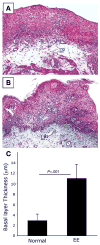
Figure 1
Figure 1
Esophageal lamina propria collagen accumulation and basal layer thickness in EE and normal individuals. A representative photomicrograph of a normal esophageal biopsy specimen shows low levels of collagen (A, original magnification, 100×). Representative photomicrograph of an EE patient’s biopsy specimen shows impressive collagen in the lamina propria and papillae (B, original magnification, 100×). The morphometric measurements of basal layer thickness in EE patients compared with normal individuals are shown in C. The data are expressed as mean ± SD. EP, epithelium; LP, lamina propria.
Figure 2
Figure 2
Collagen accumulation and basal layer thickening in the esophagus following allergen-induced EE. Representative photomicrophotograph of Masson’s trichrome-stained tissue sections of a saline-challenged mouse shows normal lamina propria collagen (A, original magnification, 100×; and B, 400×) and induced collagen accumulation in lamina propria and muscularis mucosa and extended stromal papillae following allergen challenge (C, original magnification, 100×; and D, 400×). The photomicrograph of the H&E-stained tissue section shows single or 2 cell layers thickening of basal layer in saline-challenged mice (E, original magnification, 400×) compared with 4 or 5 cell thickening in allergen-challenged mice (F, original magnification, 400×). The thickness area of basal layer is identified with the representative line in the photomicrograph (E and F). Morphometric analysis was performed to quantify basal layer and collagen thickness and is shown in G and H. The data are expressed as mean ± SD, n = 10–12 mice. EP, epithelium; LP, lamina propria; MS, muscularis mucosa; LU, lumen.
Figure 3
Figure 3
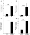
MUC5AC and TGF-β1 mRNA in the esophagus of human and experimental EE. Esophageal expression of TGF-β1 and MUC5AC mRNA was measured by performing real-time PCR analysis on normal and EE patients’ esophageal biopsy RNA (A and B) and RNA from the saline and allergen challenge WT mice (C and D). The data are expressed as mean ± SD, n = 7–9 mice/group.
Figure 4
Figure 4
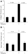
Morphometric quantification of basal layer and lamina propria collagen thickness in allergen-challenged WT and IL-5 gene-deficient mice. The basal layer and lamina propria collagen thickness in WT and IL-5 gene-deficient mice were measured by performing morphometric analysis on the esophageal tissue sections following 9 intranasal saline and allergen challenges. The basal layer thickness is shown in A and lamina propria collagen thickness in B. The data are expressed as mean ± SD, n = 12 mice.
Figure 5
Figure 5
IL-5 transgene-induced esophageal tissue remodeling. Histopathologic analysis was performed on the esophageal tissue sections of WT and IL-5 transgenic mice following Masson’s trichrome staining as shown in A–C. A representative photomicrograph of WT esophagus highlighting its basal layer thickness and lamina propria collagen level (A, original magnification, 100×). A representative photomicrograph of CD2-IL-5 transgenic mice demonstrate thickened basal layer and collagen accumulation in the lamina propria and muscularis mucosa (B, original magnification, 100×). Accumulated collagen in the lamina propria and elongated papillae is shown (C, original magnification, 400×). Morphometric analysis of basal layer thickness is shown in D, and lamina propria collagen thickness is shown in E. The data are expressed as mean ± SD, n = 12 mice. EP, epithelium; LP, lamina propria; MS, muscularis mucosa; LU, lumen.
Figure 6
Figure 6
Morphometric quantification of basal layer and lamina propria collagen thickness in allergen-challenged WT and GATA-1 gene-deficient mice. The basal layer and lamina propria collagen thickness in WT and GATA-1 gene-deficient mice were measured by performing morphometric analysis on the esophageal tissue sections following 9 intranasal saline and allergen challenges. The basal layer thickness is shown in A and lamina propria collagen thickness in B. The data are expressed as mean ± SD, n = 10–12 mice.
Figure 7
Figure 7
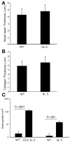
Esophageal tissue remodeling in iIL-5 transgenic mice. The levels of esophageal basal layer thickness (A) and lamina propria collagen thickness (B) of WT and iIL-5 transgenic mice are shown. The levels of eosinophils in the esophagus of CD2-IL-5 transgenic and iIL-5 transgenic mice compared with their respective control WT mice are shown in C. The data are expressed as mean ± SD, n = 12 mice.
Figure 8
Figure 8
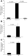
IL-5 mRNA expression in the esophagus. Esophageal expression of IL-5 mRNA was measured by performing real-time PCR analysis on the esophageal RNA from CD2-IL-5, iIL-5, and WT mice (A), saline- and allergen-challenged mice (B), and normal individuals and EE patients (C). The data are expressed as mean ± SD, n = 6–8 individuals/group.

References

    1. Bousquet J, Jeffery PK, Busse WW, et al. Asthma. From broncho-constriction to airways inflammation and remodeling. Am J Respir Crit Care Med. 2000;161:1720–1745. - PubMed
    1. Jeffery PK, Wardlaw AJ, Nelson FC, et al. Bronchial biopsies in asthma. An ultrastructural, quantitative study and correlation with hyperreactivity. Am Rev Respir Dis. 1989;140:1745–1753. - PubMed
    1. Gross TJ, Hunninghake GW. Idiopathic pulmonary fibrosis. N Engl J Med. 2001;345:517–525. - PubMed
    1. Jeffery PK. Remodeling in asthma and chronic obstructive lung disease. Am J Respir Crit Care Med. 2001;164:S28–S38. - PubMed
    1. Hoshino M, Nakamura Y, Sim JJ. Expression of growth factors and remodelling of the airway wall in bronchial asthma. Thorax. 1998;53:21–27. - PMC - PubMed

Publication types

MeSH terms