. 2007 Dec 31;179(7):1375-84.
doi: 10.1083/jcb.200705160.

Adjacent positioning of cellular structures enabled by a Cdc42 GTPase-activating protein-mediated zone of inhibition

Affiliations

Adjacent positioning of cellular structures enabled by a Cdc42 GTPase-activating protein-mediated zone of inhibition

Zongtian Tong et al. J Cell Biol. .

Abstract

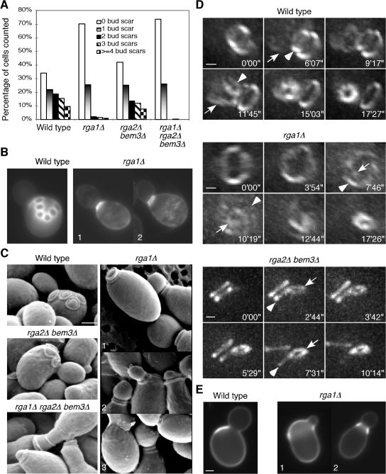
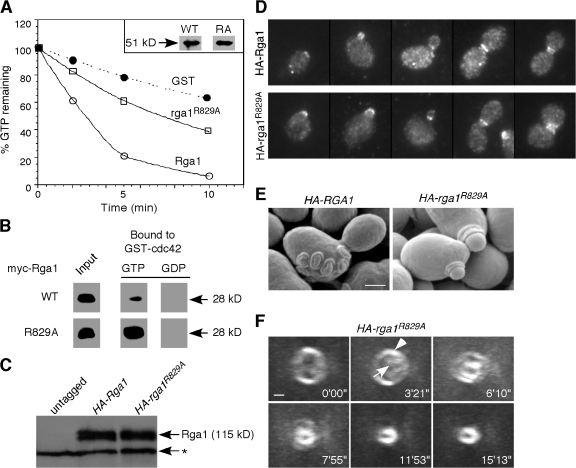
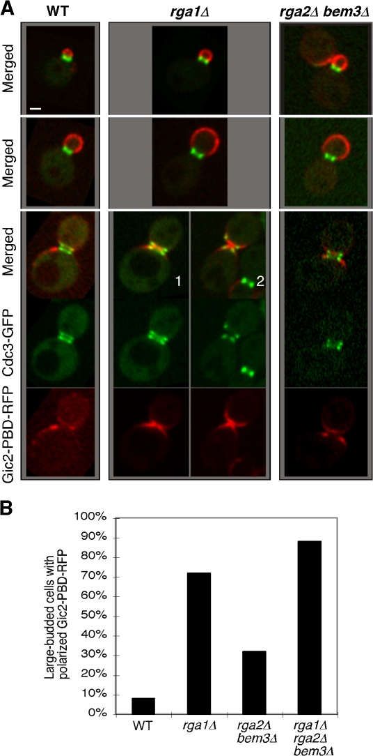
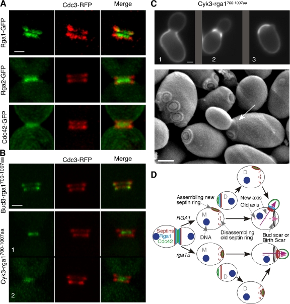
Cells of the budding yeast Saccharomyces cerevisiae are born carrying localized transmembrane landmark proteins that guide the subsequent establishment of a polarity axis and hence polarized growth to form a bud in the next cell cycle. In haploid cells, the relevant landmark proteins are concentrated at the site of the preceding cell division, to which they recruit Cdc24, the guanine nucleotide exchange factor for the conserved polarity regulator Cdc42. However, instead of polarizing at the division site, the new polarity axis is directed next to but not overlapping that site. Here, we show that the Cdc42 guanosine triphosphatase-activating protein (GAP) Rga1 establishes an exclusion zone at the division site that blocks subsequent polarization within that site. In the absence of localized Rga1 GAP activity, new buds do in fact form within the old division site. Thus, Cdc42 activators and GAPs establish concentric zones of action such that polarization is directed to occur adjacent to but not within the previous cell division site.


Figures

Figure 1.
Figure 1.
Deletion of RGA1 causes polarization and budding within the previous division site. (A) Quantitation of bud scar distribution in an asynchronous population of cells from haploid strains YEF473A (wild type), YEF2324 (rga1Δ), YEF2392 (rga2Δ bem3Δ), and YEF2380 (rga1Δ rga2Δ bem3Δ). 200 cells were counted for each strain and unbudded daughter cells were excluded. The cells with only chitin rings at the base of the growing buds were counted as having “0 bud scar.” (B) Chitin staining of wild-type and rga1Δ cells indicated in A. Double chitin rings at the neck of an rga1Δ cell (2) were visualized occasionally when the distance between the rings was large enough to be resolved by light microscopy. (C) SEM observation of bud scars. The same strains described in A were used for SEM. (D) Using the positions of the septin rings as a read-out of the budding patterns in live cells. Cells of haploid strains YZT82 (CDC3-GFP, wild type), YZT55 (rga1Δ CDC3-GFP), and YZT111 (rga2Δ bem3Δ CDC3-GFP) were grown to exponential phase in YM-P medium and observed by 3D time-lapse microscopy at 30°C. Times are given in minutes and seconds after an arbitrary starting point. Arrowheads indicate an old septin ring at the mother side of the bud neck; arrows indicate the nascent septin ring at the new bud site. Views of the 3D images from particular angles are shown: an angled side view of the bud neck of the wild-type cell, an en-face view of the mother side of the bud neck of the rga1Δ cell, and a side view of the bud neck of the rga2Δ bem3Δ cell. Please note that a clear rotation of the mother cell versus the daughter cell occurred after cytokinesis and cell separation at a time between 9 min 17 s and 11 min 45 s for the wild-type cell and between 5 min 29 s and 7 min 31 s for the rga2Δ bem3Δcell. (E) The first bud of rga1Δ daughter cells forms within the birth scar. Birth scars of representative wild-type (YEF473A) and rga1Δ (YEF2324) cells. Cells 1 and 2 represent off-center and central budding within the birth scar, respectively. Bars, 1 μm.
Figure 2.
Figure 2.
The role of Rga1 in polarity axis determination depends on its Cdc42-GAP activity. (A) GAP assays. Cdc42 prebound to γ-[32P]GTP was incubated with GST, the GST-Rga1 GAP domain, or the same domain containing the R829A mutation, and radioactivity remaining bound to Cdc42 is plotted against time of incubation. The inset shows that similar amounts of wild-type and mutant GAP domains were used in the assay. This GAP assay is representative of three experiments with consistent results. (B) Binding assays. Recombinant myc-tagged Rga1 or rga1R829A GAP domains were incubated with bead-bound recombinant GST-cdc42Q61L (GTP-bound) or GST-cdc42T17N (GDP-bound) to assess binding. (C) HA-Rga1 and HA-rga1R829A are expressed at similar levels. Protein samples were prepared from YZT194 (HA-RGA1) and YZT195 (HA-rga1R829A). The asterisk indicates a cross-reacting protein with the HA-antibody in yeast extracts. (D) HA-Rga1 and HA-rga1R829A display similar localization patterns in the cell cycle. Strains YZT194 and YZT195 were grown to exponential phase in YM-P medium at 23°C and examined by immunofluorescence using an anti-HA antibody. (E) SEM observation of bud scars of strains YZT194 and YZT195. (F) Visualization of new septin ring formation in live cells of strain YZT198 (HA-rga1R829A CDC3-GFP) by 3D time-lapse microscopy. Eight out of nine cells were observed to form a new septin ring within the old ring. The arrowhead indicates the old septin ring; the arrow indicates the new septin ring. Bars, 1 μm.
Figure 3.
Figure 3.
Deletion of RGA1 causes an elevated level of GTP-Cdc42 at the cell division site. (A) Cells of YZT292 (WT), YZT293 (rga1Δ), and YZT294 (rga2Δ bem3Δ) carrying integrated CDC3-GFP and GIC2-PBD-RFP were imaged by two-color light microscopy. Single representative GFP and RFP images from a stack of z sections for each cell were selected to show the localization patterns of Cdc3-GFP and Gic2-PBD-RFP with high resolution. Bar, 1 μm. (B) Quantitation of large-budded cells with neck-localized Gic2-PBD-RFP. Cells of YZT295 (rga1Δ rga2Δ bem3Δ) and other strains as in A were used and only large-budded cells with a clear septum (n = 50 for each strain) were scored.
Figure 4.
Figure 4.
Targeting of the Rga1 GAP domain to the division site by heterologous proteins during and after cytokinesis is sufficient for the role of Rga1 in polarity-axis determination. (A) Fine patterns of Rga1, Rga2, and Cdc42 localization with respect to the septin rings during cytokinesis. Live cells of YZT211 (RGA1-GFP CDC3-DsRed), YZT166 (CDC3-mCherry) carrying plasmid pRS426-RGA2-GFP (the endogenous level of Rga2 was difficult to detect, thus, a high-copy plasmid carrying Rga2-GFP was used here; the pattern of Rga2 localization did not change with the high-copy plasmid, but the GFP signal was significantly improved), and YZT221 (CDC42-GFP CDC3-DsRed) were observed by 3D fluorescence microscopy at 23°C. All images in A and B are oriented such that the bud side is up. (B) Localization of Bud3-Rga1-GAP and Cyk3-Rga1-GAP fusions during cytokinesis. Cells of YZT240 (BUD3-rga1700–1007aa-GFP CDC3-mCherry) and YZT241 (CYK3-rga1700–1007aa-GFP CDC3-mCherry) were observed by 3D fluorescence microscopy as described for A. (C) Differential suppression of budding pattern defects by the Cyk3-Rga1-GAP fusion for the mother and daughter cells after cell division. (top) Birth scars of the strain JGY1645 (rga1Δ CYK3-rga1700–1007aa-GFP) were visualized. 1 and 2 represent off-center and central budding within the birth scar, respectively; 3 represents axial budding in a suppressed cell. (bottom) Birth scar (arrow) and bud scars of the strain JGY1645 were visualized by SEM. Strain JGY1645 was grown to exponential phase in YP medium containing 2% glycerol (YPG) and processed for SEM. The birth scar appeared to be better visualized in the YPG-poor medium than in the YPD-rich medium. Only 3.3% of the CYK3-RGA1-GAP mother cells (n = 273) budded within the bud scar when the strain was grown in YPG medium. (D) A model for the role of Rga1 in polarity-axis determination. New axis and Old axis refer to the axes of polarized cell growth. M, mother; D, daughter. Bars, 1 μm.

References

    1. Ahmadian, M.R., P. Stege, K. Scheffzek, and A. Wittinghofer. 1997. Confirmation of the arginine-finger hypothesis for the GAP-stimulated GTP-hydrolysis reaction of Ras. Nat. Struct. Biol. 4:686–689. - PubMed
    1. Barton, A.A. 1950. Some aspects of cell division in Saccharomyces cerevisiae. J. Gen. Microbiol. 4:84–86. - PubMed
    1. Bender, A., and J.R. Pringle. 1989. Multicopy suppression of the cdc24 budding defect in yeast by CDC42 and three newly identified genes including the ras–related gene RSR1. Proc. Natl. Acad. Sci. USA. 86:9976–9980. - PMC - PubMed
    1. Bernards, A. 2003. GAPs galore! A survey of putative Ras superfamily GTPase activating proteins in man and Drosophila. Biochim. Biophys. Acta. 1603:47–82. - PubMed
    1. Bernards, A., and J. Settleman. 2004. GAP control: regulating the regulators of small GTPases. Trends Cell Biol. 14:377–385. - PubMed

Publication types

MeSH terms

Substances