. 2007 Dec 31;179(7):1497-510.
doi: 10.1083/jcb.200708107.

Molecular basis of synaptic vesicle cargo recognition by the endocytic sorting adaptor stonin 2

Affiliations

Molecular basis of synaptic vesicle cargo recognition by the endocytic sorting adaptor stonin 2

Nadja Jung et al. J Cell Biol. .

Abstract

Synaptic transmission depends on clathrin-mediated recycling of synaptic vesicles (SVs). How select SV proteins are targeted for internalization has remained elusive. Stonins are evolutionarily conserved adaptors dedicated to endocytic sorting of the SV protein synaptotagmin. Our data identify the molecular determinants for recognition of synaptotagmin by stonin 2 or its Caenorhabditis elegans orthologue UNC-41B. The interaction involves the direct association of clusters of basic residues on the surface of the cytoplasmic domain of synaptotagmin 1 and a beta strand within the mu-homology domain of stonin 2. Mutation of K783, Y784, and E785 to alanine within this stonin 2 beta strand results in failure of the mutant stonin protein to associate with synaptotagmin, to accumulate at synapses, and to facilitate synaptotagmin internalization. Synaptotagmin-binding-defective UNC-41B is unable to rescue paralysis in C. elegans stonin mutant animals, suggesting that the mechanism of stonin-mediated SV cargo recognition is conserved from worms to mammals.


Figures

Figure 1.
Figure 1.
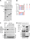
The synaptotagmin 1 C2A domain is required for stonin 2–mediated synaptotagmin 1 internalization. (A) Stonin 2 recruits synaptotagmin 1 to AP-2–coated puncta. HEK293 cells stably expressing FLAG–synaptotagmin 1 were subjected to anti-FLAG antibody uptake experiments. Internalized synaptotagmin 1 (green) colocalizes partially with endogenous AP-2 (red) in plasmalemmal puncta (yellow), presumably representing clathrin/AP-2–coated pits. Blue, DAPI-stained nuclei. Images were obtained by deconvolution fluorescence microscopy. White boxes indicate magnified areas below. Bars: (top) 10 μm; (bottom) 3 μm. (B) HEK293 cells coexpressing HA-tagged stonin 2 and FLAG–synaptotagmin 1 C2 domain deletion mutants were analyzed by antibody uptake assays as described in A. Surface-exposed (red) or internalized (green) synaptotagmin 1 was labeled with Alexa Fluor594– or 488–conjugated secondary antibodies, respectively. Blue, DAPI-stained nuclei. Equal exposure times and identical intensity normalization were used during the acquisition of the images. Bars, 20 μm. (C) The C2A domain is required for membrane recruitment of stonin 2. NIE cells were cotransfected with AP-2–binding–deficient HA-stonin 2δWFδNPF and FLAG–synaptotagmin 1 deletion constructs. The distribution of surface synaptotagmin 1 (red) and stonin 2 (green) were analyzed by deconvolution fluorescence microscopy. Blue, DAPI-stained nuclei. White boxes indicate magnified areas to the right. Bars, 20 μm. (D) Deletion of the C2A domain results in loss of stonin 2 binding in coimmunoprecipitation experiments. HEK293 cells were cotransfected with HA-tagged stonin 2 and FLAG-tagged synaptotagmin 1 deletion constructs. Immunoprecipitation was performed using antibodies directed against the FLAG tag. Immunoprecipitated proteins were detected by immunoblotting for clathrin heavy chain (CHC) as control, HA-tag (stonin 2), α-adaptin (AP-2α), and FLAG-tag (syt1). 3% of the input material was loaded as standard.
Figure 2.
Figure 2.
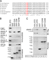
Clusters of basic amino acids within the synaptotagmin 1 C2 domains are involved in the association with stonin 2. (A) Stonin 2 and synaptotagmin 1–C2A interact directly. Stonin 2–His6 was purified from stably transfected HEK293 cells and incubated with GST–synaptotagmin 1 fusion proteins immobilized on beads. Bound stonin 2 was detected by immunoblotting for the His6-tag. Arrows mark bound stonin 2 on the Ponceau-stained nitrocellulose membrane. 10% of the input was loaded as standard (STD). Molecular masses are indicated. (B) Alignment of basic patches within synaptotagmin 1 C2A and C2B domains and mutants analyzed. (C) GST-C2A WT or mutant fusion proteins (see B) were assayed in pulldown experiments for their ability to associate with HA–stonin 2δWFδNPF from HEK293 lysates. Samples were analyzed by immunoblotting for HA–stonin 2, AP-2β, and clathrin heavy chain (CHC). 7% of the input was loaded as standard. (D) Coimmunoprecipitation experiment performed from cotransfected fibroblasts. HEK293 cells were cotransfected with HA-tagged stonin 2 and FLAG-tagged synaptotagmin 1 WT and mutant (K189–191E; K213E; K244E; KR321, 322EE; and K324–327EE) constructs. Immunoprecipitation was performed using polyclonal antiserum against stonin 2. Immunoblotting was performed using antibodies against HA-stonin 2, AP-2β, anti–FLAG–synaptotagmin 1, and anti–clathrin heavy chain antibodies. 3% of the input was loaded as standard. Splices are indicated by solid lines (all taken from the same exposure). In the case of FLAG–synaptotagmin 1, the bands representing material present in 3% of the starting material were taken from a slightly longer exposure of the x-ray film from the same immunoblot to reveal the bands. The two exposures are indicated by black boxes. Black lines indicate that intervening lanes have been spliced out.
Figure 3.
Figure 3.
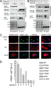
Evolutionarily conserved residues within a β strand of its μHD are required for association of stonin 2 with synaptotagmin 1. (A) The KYE site is evolutionarily conserved. Multiple protein sequence alignment of stonin 2 and orthologues from rat, mouse, human, cattle, zebrafish, D. melanogaster, and C. elegans. Numbers refer to the last amino acid residue within the predicted β strand. Conserved residues K783, Y784, and E785 are colored in red. (B) Mutation of conserved residues K783A, Y784A, and E785A within stonin 2 abolishes its ability to bind to synaptotagmin 1. GST–synaptotagmin 1 fusion proteins immobilized on beads were incubated with cell extracts derived from HEK293 cells transfected with AP-2–binding–deficient HA–stonin 2δWFδNPF, HA–stonin 2δKYE, or HA–stonin 2δWFδNPFδKYE, respectively. Bound proteins were detected by immunoblotting for clathrin heavy chain (CHC), HA–stonin 2, and AP-2μ. 8% of the input was loaded as standard (STD). (C) In vitro binding experiment using purified recombinant proteins. GST-C2AB immobilized on beads was incubated with purified stonin 2–His6 WT or δKYE. Bound stonin 2–His6 was detected by immunoblotting for the His6-tag. The arrow marks the stonin 2–His6 band on the Ponceau S–stained nitrocellulose membrane. 10% of the input was loaded as standard.
Figure 4.
Figure 4.
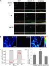
Stonin 2–mediated synaptotagmin 1 internalization requires an intact KYE site within stonin 2. (A) Synaptotagmin 1 does not associate with mutant stonin 2δKYE in coimmunoprecipitation experiments. HEK293 cells were cotransfected with FLAG–synaptotagmin 1 and HA–stonin 2WT, AP-2–binding–deficient HA–stonin 2δWFδNPF, or HA–stonin 2δKYE, respectively. Proteins immunoprecipitated with antibodies against stonin 2 were detected by immunoblotting for HA–stonin 2, AP-2β, FLAG–synaptotagmin 1, and clathrin heavy chain (CHC). 2% of the input was loaded as standard. (B) The Y784R point mutant of stonin 2 is unable to associate with synaptotagmin 1. Experiments were done as described in A. 2.5% of the input was loaded as standard. (C) Stonin 2δKYE and stonin 2YR mutants lack the ability to facilitate synaptotagmin 1 internalization. HEK-syt1 cells were transfected with HA-tagged stonin 2WT, stonin 2δKYE, or stonin 2YR, respectively. Synaptotagmin 1 internalization was assayed as described in the Fig. 1 legend. Red, surface synaptotagmin 1; green, internal synaptotagmin 1; blue, DAPI-stained nuclei. Stonin 2 expression was verified using Cy5-labeled secondary antibodies (right). Equal exposure times and identical intensity normalization were used during image acquisition. Bars, 20 μm. (D) Quantifications of synaptotagmin internalization experiments using stonin 2 mutants. Fluorescence intensities were quantified by applying the Mask function of the Slidebook 4.0.8 Digital Microscopy Software (Intelligent Imaging Innovations) on the green channel (internalized synaptotagmin 1) in stonin 2–transfected cells (red). Error bars represent SEM.
Figure 5.
Figure 5.
The stonin 2 KYE site is required for synaptotagmin 1–dependent recruitment of stonin 2 to the plasmalemma in neuroblastoma cells. NIE cells were cotransfected with lumenally FLAG-tagged synaptotagmin 1 and HA-tagged stonin 2WT (A), AP-2–binding–deficient stonin 2δWFδNPF (B), stonin 2δKYE (C), or stonin 2YR (D), respectively. Surface synaptotagmin 1 was labeled by anti-FLAG antibodies (red). After permeabilization, stonin 2 was immunostained using antibodies directed against stonin 2 (green). White boxes indicate magnifications to the right. Bars, 20 μm.
Figure 6.
Figure 6.
The stonin 2 KYE site is required for synaptic localization of stonin 2 and facilitates targeting of synaptotagmin 1 to recycling vesicles in primary neurons. (A) Primary hippocampal neurons at DIV9 were transfected with HA–stonin 2WT, –stonin 2YR, or –stonin 2δKYE, respectively. Monoclonal antibodies against synaptotagmin 1 are used to decorate presynaptic sites (red). Low-power views (top) exemplify the overall distribution of HA-tagged stonin 2 or mutants (green; insets, threefold magnified images of boxed area). High-power views (bottom) represent the localization of HA–stonin 2 or mutants in selected neurites. Bars, 10 μm. (B–D) Stonin 2WT but not stonin 2δKYE mutant enhances targeting of sytpHluorin to recycling vesicles in primary hippocampal neurons. (B) To assess the surface and vesicular pools of sytpHluorin, the relative fluorescence (F) values were measure after acid quenching and ammonium dequenching. Bar, 10 μm. (C) Quantitative analysis of n = 7 experiments, each comprising >50 boutons as in B. Mean time course of sytpHluorin at synaptic boutons normalized to initial F values is shown. (D) Ratios of vesicular/surface stranded pools of sytpHluorin (n = 7 experiments; error bars represent SEM; control, 3.05 ± 0.3; stonin 2WT, 4.28 ± 0.19; stonin 2δKYE, 2.6 ± 0.15; **, P < 0.01). Expression of WT stonin 2 results in a significant increase in the vesicular/surface pool ratio, whereas stonin 2δKYE has lost this ability.
Figure 7.
Figure 7.
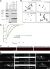
The KYE site is required for UNC-41 function in locomotory behavior in C. elegans. (A) Mutation of the KYE site within C. elegans UNC-41B abolishes its ability to bind to synaptotagmin 1. GST or GST fused to the C2 domains of rat or C. elegans (Ce) synaptotagmin 1 immobilized on 30-μg protein beads were incubated with cell extracts derived from Cos7 cells transfected with HA–UNC-41B or HA–UNC-41B δKYE. Bound proteins were detected by immunoblotting for HA–UNC-41B, actin, and AP-2μ. 4% of the input was loaded as standard. (B and C) Worm tracking assay for unc-41 mutant (unc-41) and rescued animals. (B) Representative worm moving traces over 10 min. The number of worms on each plate is indicated. (C) The mean speed for each 2 s was used and the percentage of the speed below a certain threshold was plotted for each genotype. Data are given as mean ± SEM. (D) Confocal images of immobilized worms expressing endogenous UNC-41 or plasmid-borne UNC-41B WT or the δKYE mutant analyzed by indirect immunofluorescence microscopy using UNC-41–specific antibodies. Bar, 10 μm. (E) Confocal images of immobilized worms expressing GFP–UNC-41B WT or the δKYE mutant. A magnified view of the boxed area from the dorsal cord is shown to the right of the image. Bar, 10 μm.

Similar articles

Cited by

References

    1. Bai, J., W.C. Tucker, and E.R. Chapman. 2004. PIP2 increases the speed of response of synaptotagmin and steers its membrane-penetration activity toward the plasma membrane. Nat. Struct. Mol. Biol. 11:36–44. - PubMed
    1. Bennett, M.K., N. Calakos, T. Kreiner, and R.H. Scheller. 1992. Synaptic vesicle membrane proteins interact to form a multimeric complex. J. Cell Biol. 116:761–775. - PMC - PubMed
    1. Bonifacino, J.S., and L.M. Traub. 2003. Signals for sorting of transmembrane proteins to endosomes and lysosomes. Annu. Rev. Biochem. 72:395–447. - PubMed
    1. Brodin, L., P. Low, and O. Shupliakov. 2000. Sequential steps in clathrin-mediated synaptic vesicle endocytosis. Curr. Opin. Neurobiol. 10:312–320. - PubMed
    1. Chapman, E.R., R.C. Desai, A.F. Davis, and C.K. Tornehl. 1998. Delineation of the oligomerization, AP-2 binding, and synprint binding region of the C2B domain of synaptotagmin. J. Biol. Chem. 273:32966–32972. - PubMed

Publication types

MeSH terms