

Progressive aphasia secondary to Alzheimer disease vs FTLD pathology
- PMID: 18166704
- PMCID: PMC2749307
- DOI: 10.1212/01.wnl.0000287073.12737.35
Progressive aphasia secondary to Alzheimer disease vs FTLD pathology
Abstract
Background: The pathology causing progressive aphasia is typically a variant of frontotemporal lobar degeneration, especially with ubiquitin-positive inclusions (FTLD-U). Less commonly the underlying pathology is Alzheimer disease (AD).
Objective: To compare clinicopathologic and MRI features of subjects with progressive aphasia and AD pathology to subjects with aphasia and FTLD-U pathology and subjects with typical AD.
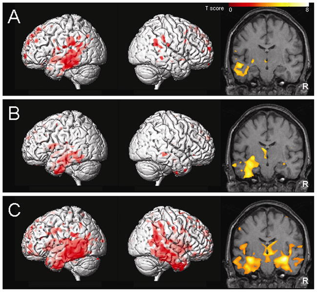
Methods: We identified 5 subjects with aphasia and AD pathology and 5 with aphasia and FTLD-U pathology with an MRI from a total of 216 aphasia subjects. Ten subjects with typical AD clinical features and AD pathology were also identified. All subjects with AD pathology underwent pathologic reanalysis with TDP-43 immunohistochemistry. Voxel-based morphometry (VBM) was used to assess patterns of gray matter atrophy in the aphasia cases with AD pathology, aphasia cases with FTLD-U, and typical AD cases with AD pathology, compared with a normal control group.
Results: All aphasic subjects had fluent speech output. However, those with AD pathology had better processing speed than those with FTLD-U pathology. Immunohistochemistry with TDP-43 antibodies was negative. VBM revealed gray matter atrophy predominantly in the temporoparietal cortices, with notable sparing of the hippocampus in the aphasia with AD subjects. In comparison, the aphasic subjects with FTLD-U showed sparing of the parietal lobe. Typical AD subjects showed temporoparietal and hippocampal atrophy.
Conclusions: A temporoparietal pattern of atrophy on MRI in patients with progressive fluent aphasia and relatively preserved processing speed is suggestive of underlying Alzheimer disease pathology rather than frontotemporal lobar degeneration with ubiquitin-only immunoreactive changes.
Figures
References
-
- Mesulam MM. Slowly progressive aphasia without generalized dementia. Ann Neurol. 1982;11:592–598. - PubMed
-
- Neary D, Snowden JS, Gustafson L, et al. Frontotemporal lobar degeneration: a consensus on clinical diagnostic criteria. Neurology. 1998;51:1546–1554. - PubMed
-
- Kertesz A, Davidson W, McCabe P, et al. Primary progressive aphasia: diagnosis, varieties, evolution. J Int Neuropsychol Soc. 2003;9:710–719. - PubMed
Publication types
MeSH terms
Substances
Grants and funding
LinkOut - more resources
Full Text Sources
Medical