Review
. 2008 Jan;18(1):48-63.
doi: 10.1038/cr.2008.7.

XRCC1 and DNA polymerase beta in cellular protection against cytotoxic DNA single-strand breaks

Affiliations
Review

XRCC1 and DNA polymerase beta in cellular protection against cytotoxic DNA single-strand breaks

Julie K Horton et al. Cell Res. 2008 Jan.

Abstract

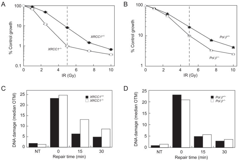
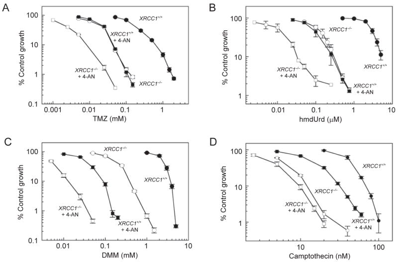
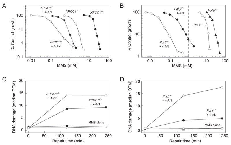
Single-strand breaks (SSBs) can occur in cells either directly, or indirectly following initiation of base excision repair (BER). SSBs generally have blocked termini lacking the conventional 5'-phosphate and 3'-hydroxyl groups and require further processing prior to DNA synthesis and ligation. XRCC1 is devoid of any known enzymatic activity, but it can physically interact with other proteins involved in all stages of the overlapping SSB repair and BER pathways, including those that conduct the rate-limiting end-tailoring, and in many cases can stimulate their enzymatic activities. XRCC1(-/-) mouse fibroblasts are most hypersensitive to agents that produce DNA lesions repaired by monofunctional glycosylase-initiated BER and that result in formation of indirect SSBs. A requirement for the deoxyribose phosphate lyase activity of DNA polymerase beta (pol beta) is specific to this pathway, whereas pol beta is implicated in gap-filling during repair of many types of SSBs. Elevated levels of strand breaks, and diminished repair, have been demonstrated in MMS-treated XRCC1(-/-), and to a lesser extent in pol beta(-/-) cell lines, compared with wild-type cells. Thus a strong correlation is observed between cellular sensitivity to MMS and the ability of cells to repair MMS-induced damage. Exposure of wild-type and pol beta(-/-) cells to an inhibitor of PARP activity dramatically potentiates MMS-induced cytotoxicity. XRCC1(-/-) cells are also sensitized by PARP inhibition demonstrating that PARP-mediated poly(ADP-ribosyl)ation plays a role in modulation of cytotoxicity beyond recruitment of XRCC1 to sites of DNA damage.


Figures

Figure 1
Figure 1
Survival of mouse embryonic fibroblasts following exposure to IR and repair of IR-induced DNA damage. (A) XRCC1/ (closed circles) and XRCC1/ (open circles) and (B) Pol β +/+ (closed triangles) and pol β / cells (open triangles) were exposed to IR and survival was determined by growth inhibition assays as described [119]. The dashed line indicates the dose of IR used in the experiments presented in panels C and D. Data are from representative experiments where the values are the mean ± SEM of 4–5 determinations. (C and D) DNA damage (median OTM) was analyzed in non-treated cells (NT), and cells irradiated (5 Gy) then allowed to repair for 15 and 30 min as shown. The XRCC1 [73] and pol β [26] cell lines have been described previously.
Figure 2
Figure 2
Complementation of XRCC1/ hypersensitivity phenotypes by expression of the XRCC1 gene. XRCC1+/+ (closed circles) and XRCC1/ mouse embryonic fibroblasts (open circles) or XRCC1/ fibroblasts transfected with mouse XRCC1 cDNA (closed diamonds) were treated with (A) MMS for 1 h, (B) TMZ for 4 h, (C) hmdUrd for 24 h, or (D) camptothecin for 24 h. Cell sensitivity was determined by growth inhibition assays as described [119]. The cell lines have been described previously [73].
Figure 3
Figure 3
MMS hypersensitivity in XRCC1/ compared with pol β / mouse embryonic fibroblasts. Sensitivity of XRCC1/ (open circles) and pol β / cells (open triangles) to a 1 h exposure to MMS is compared with that of the respective isogenic wild-type (+/+) cell lines (closed symbols). Cell sensitivity was determined by growth inhibition assays as described [119]. The cell lines have been described previously [26, 73]. Data taken from ref. [106].
Figure 4
Figure 4
Increased DNA damage following exposure of repair-deficient mouse embryonic fibroblasts to MMS. (A and B) DNA damage (median OTM) after treatment of cells (as indicated) for 20 min with 10 mM MMS in ice-cold medium, and then during repair in warm medium for up to 120 min. NT are non-treated cells. (C) Sensitivity of XRCC1+/+ (closed circles) and XRCC1/ (open circles) and (D) Pol β+/+ (closed triangles), pol β / (open triangles) cells to a 20 min exposure to MMS in ice-cold medium. Growth inhibition assays were conducted as described [119]. Data are from representative experiments where the values are the mean ± SEM of 3 independent determinations. The dashed line indicates the concentration of cold MMS used in the experiments presented in panels A and B.
Figure 5
Figure 5
XRCC1- and pol β-dependent hypersensitivity to monofunctional mustards. XRCC1+/+ (closed circles) and XRCC1/ (open circles) mouse embryonic fibroblasts were treated with (A) DMM or (B) DEM for 1 h. (C) Pol β+/+ (closed symbols) and pol β / (open symbols) cells were treated with DMM for 1 h in the presence (squares) or absence (triangles) of pamoic acid (PA) (pretreatment with 300 μM for 7 h, then continued incubation following DMM for a total of 24 h). Data represent the mean ± SEM of at least 3 independent experiments. Cell sensitivity was determined by growth inhibition assays as described [119].
Figure 6
Figure 6
Hypersensitivity of XRCC1/ mouse embryonic fibroblasts to DNA damaging agents and effect of PARP inhibition. XRCC1+/+ (closed symbols) and XRCC1/ cells (open symbols) were treated with (A) TMZ for 4 h, (B) hmdUrd for 24 h, (C) DMM for 1 h or (D) camptothecin for 24 h. Shown are survival curves for ‘control’ cells in the absence of PARP inhibitor (circles), or cells treated in the presence of 4-AN (5 μM for 24 h) (squares). Data represent the mean ± SEM of at least 3 independent experiments. Cell sensitivity was determined by growth inhibition assays as described [119].
Figure 7
Figure 7
Enhanced cytotoxicity and increased DNA damage following co-exposure to MMS and 4-AN. (A) XRCC1+/+ (closed symbols) and XRCC1/ (open symbols) and (B) Pol β+/+ (closed symbols) and pol β / (open symbols) mouse embryonic fibroblasts were treated with MMS in ice-cold medium for 20 min. Shown are survival curves for ‘control’ cells in the absence of PARP inhibitor (circles or triangles), or cells treated in the presence of 4-AN for a total of 24 h (squares or diamonds). Cell sensitivity was determined by growth inhibition assays as described [119]. Data are from a representative experiment where the values are the mean of triplicate determinations. The dashed line indicates the concentration of cold MMS combined with 4-AN used in the experiments presented in panels C and D. (C) XRCC1+/+ and XRCC1/and (D) Pol β+/+ and pol β /cells were treated for 20 min with 1 mM MMS in ice-cold medium in the absence or presence of 4-AN (symbols as described for panels A and B). Plotted is DNA damage (median OTM) during cellular repair in warm medium ± 4-AN for up to 4 h.

References

    1. Lindahl T. Instability and decay of the primary structure of DNA. Nature. 1993;362:709–715. - PubMed
    1. Srivastava DK, Vande Berg BJ, Prasad R, et al. Mammalian abasic site base excision repair. Identification of the reaction sequence and rate-determining steps. J Biol Chem. 1998;273:21203–21209. - PubMed
    1. Sossou M, Flohr-Beckhaus C, Schulz I, et al. APE1 overexpression in XRCC1-deficient cells complements the defective repair of oxidative single strand breaks but increases genomic instability. Nucleic Acids Res. 2005;33:298–306. - PMC - PubMed
    1. Ward JF. DNA damage produced by ionizing radiation in mammalian cells: identities, mechanisms of formation, and reparability. Prog Nucleic Acid Res Mol Biol. 1988;35:95–125. - PubMed
    1. Izumi T, Hazra TK, Boldogh I, et al. Requirement for human AP endonuclease 1 for repair of 3′Œ-blocking damage at DNA singlestrand breaks induced by reactive oxygen species. Carcinogenesis. 2000;21:1329–1334. - PubMed

Publication types

MeSH terms