Comparative Study
. 2008 Mar;99(3):486-96.
doi: 10.1111/j.1349-7006.2007.00702.x. Epub 2007 Dec 27.

Involvement of protein kinase C beta-extracellular signal-regulating kinase 1/2/p38 mitogen-activated protein kinase-heat shock protein 27 activation in hepatocellular carcinoma cell motility and invasion

Affiliations
Comparative Study

Involvement of protein kinase C beta-extracellular signal-regulating kinase 1/2/p38 mitogen-activated protein kinase-heat shock protein 27 activation in hepatocellular carcinoma cell motility and invasion

Kun Guo et al. Cancer Sci. 2008 Mar.

Abstract

To understand the molecular mechanism that underlies the role of various prominent signal pathways in hepatocellular carcinoma (HCC) metastasis, a human signal transduction oligonucleotide microarray analysis was carried out in cultured HCC cell models with increasing spontaneous metastatic potential (MHCC97L, MHCC97H, and HCCLM6). The results revealed that the mitogen-activated protein kinase (MAPK) pathway is the prominently upregulated pathway in HCC metastasis. Further study showed that basal phosphorylated levels of extracellular signal-regulating kinase (ERK)(1/2) and p38 MAPK consecutively increased from MHCC97L to MHCC97H to HCCLM6 cells, but not c-Jun N-terminal kinase. The phosphorylation of ERK(1/2) and p38 MAPK was regulated by upregulated protein kinase C beta (PKC beta) in HCC cells through the integrated use of PKC beta RNA interference, the PKC beta specific inhibitor enzastaurin and a PKC activator phorbol-12-myristate-13-acetate. Heat shock protein 27 (HSP27) was also verified as a downstream common activated protein of PKC beta-ERK(1/2) and PKC beta-p38 MAPK. In vitro migration and invasion assay further showed that the depletion of PKC beta or inhibition of PKC beta activation effectively decreased HCC cell motility and invasion. Moreover, the motility and invasion of phorbol-12-myristate-13-acetate-stimulated PKC beta-mediated HCC cells was significantly negated by an ERK inhibitor, 1.4-diamino-2.3-dicyano-1.4-bis[2-aminophenylthio] butadiene, or a p38 MAPK inhibitor, 4-(4-Fluorophenyl)-2-(4-methylsulfinylphenyl)-5-(4-pyridyl)1H-imidazole. It also showed that HSP27 is critical in PKC beta-mediated HCC cell motility and invasion. Taken together, this study reveals the important role of this PKC beta-ERK(1/2)/p38MAPK-HSP27 pathway, which was verified for the first time, in modulating HCC cell motility and invasion.


Figures
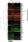
Figure 1
Figure 1
Clustering analysis of differently expressed genes in three hepatocellular carcinoma cell lines. Hierarchical clustering was carried out using Pearson's correlation distance and averaging linkage of cell lines and genes. Each row represents one of 62 differently expressed genes; each column represents a separate swap‐dye experiment. High and low expression are shown in red and green, respectively. –R, reverse‐dye experiment.
Figure 2
Figure 2
The extracellular signal‐regulating kinase (ERK)1/2 pathway, p38 mitogen‐activated protein kinase (MAPK) pathway, and protein kinase Cβ (PKCβ) were consecutively activated in metastatic hepatocellular carcinoma cell clones MHCC97L, MHCC97H, and HCCLM6. The levels of ERK1/2, p38 MAPK, c‐Jun N‐terminal kinase, and PKCβ and their basal phosphorylation were determined by immunoblot analysis of whole cell lysate (30 µg) using specific and phosphospecific antibodies. The values on top of the bands represent the densitometric estimation of the relative density of the band, calculated by comparing the ratio of phosphor‐protein versus total protein intensity of MHCC97H and HCCLM6 cell lines with that of MHCC97L cell line (the ratio in MHCC97L was set as baseline 1.0). The level of glyceraldehyde‐3‐phosphate dehydrogenase served as the loading control.
Figure 3
Figure 3
Extracellular signal‐regulating kinase (ERK)1/2 and p38 mitogen‐activated protein kinase (MAPK) pathways were efficiently activated by upstream protein kinase Cβ (PKCβ) in hepatocellular carcinoma (HCC) cells. (a) PKC inhibition assays with a series concentration of enzastaurin (LY317615; 0, 0.025, 0.05, 0.1, 1, 2.5, and 10 µM) were carried out in HCC cells the using HTScan PKC Kinase Assay Kit from Cell Signaling Technology (Beverly, MA). Inhibition of PKCα, PKCβ, PKCγ, or PKCδ by various concentrations of LY317615 was determined through detecting fluorescence values of reaction solutions. The fluorescence value of reaction solution with 0 µM LY317615 served as a percent control for each PKC inhibition assay. (b) HCCLM6 cells pretreated with 0.025 µM LY317615 for 1.5 h and the cell extracts (20 µg) were subjected to sodium dodecyl sulfate–polyacrylamide gel electrophoresis (SDS‐PAGE), the levels of ERK1/2, p38 MAPK, c‐Jun N‐terminal kinase, heat shock protein 27 (HSP27), and their phosphorylation were determined by immunoblot analysis. The values underneath the bands represent the densitometric estimation of the relative density of the band, calculated by comparing the ratio of phosphor‐protein versus total protein intensity of LY317615‐treated HCCLM6 cells with that of HCCLM6 cells without LY317615 treatment (the ratio in HCCLM6 cells without LY317615 treatment was set as baseline 1.0). Similar findings were also observed in metastatic HCC cell clones MHCC97L and MHCC97H (data not shown). (c) HCCLM6 cells were transfected with PKCβ small interfering RNA (siRNA)‐1 (70 nM), siRNA‐2 (70 nM), siRNA‐3 (70 nM), and negative control (non‐specific siRNA) for 48 h. Equal amounts of cell extracts (30 µg) were subjected to SDS‐PAGE, and the levels of total PKCβ were determined by immunoblot analysis. The level of glyceraldehyde‐3‐phosphate dehydrogenase (GAPDH) served as the loading control. The values underneath the bands represent the densitometric estimation of the relative density of the band, calculated by comparing the ratio of PKCβversus GAPDH with that of HCCLM6 cells without siRNA treatment (the ratio in HCCLM6 cells without siRNA treatment was set as baseline 1.0). (d) After HCCLM6 cells were transfected with PKCβ siRNA‐1 (70 nM) followed by phorbol‐12‐myristate‐13‐acetate (20 nM) for 4 h, levels of phosphor‐ERK1/2, phosphor‐p38 MAPK, phosphor‐HSP27, and total proteins were estimated. The values underneath the bands represent the relative ratios of phosphorylation versus total protein compared with that of HCCLM6 cells. Similar findings were also observed in MHCC97L and MHCC97H cells (data not shown).
Figure 4
Figure 4
Phosphorylation of heat shock protein 27 (HSP27) was regulated by protein kinase Cβ (PKCβ)–extracellular signal‐regulating kinase (ERK)1/2 and PKCβ–p38 mitogen‐activated protein kinase (MAPK) in hepatocellular carcinoma (HCC) cells. (a) Expression of total HSP27 was analyzed using the BioSource Human HSP27 kit. Equal amounts of total protein, each at a final concentration of 50 µg/mL, from metastatic HCC cell clones MHCC97L, MHCC97H, and HCCLM6 were analyzed using the BioSource Human HSP27 kit and expression of total HSP27 was consecutively increased. Basal phosphorylated level of HSP27 was detected by immunoblot analysis in three HCC cell lines. The values on top of the bands represent the relative ratio of basal phosphorylation in MHCC97L, MHCC97H, and HCCLM6 cells. (b) HCCLM6 cells were treated with 4‐(4‐Fluorophenyl)‐2‐(4‐methylsulfinylphenyl)‐5‐(4‐pyridyl)1H‐imidazole (SB203580; 5 µM) for 2 h, 1.4‐diamino‐2.3‐dicyano‐1.4‐bis[2‐aminophenylthio] butadiene (U0126; 10 µM) for 1.5 h, or 1,9‐pyrazoloanthrone (SP600125; 10 µM) for 2 h. Expression of total ERK1/2, p38 MAPK, c‐Jun N‐terminal kinase, HSP27 and their phosphorylated forms were analyzed using specific antibodies by immunoblot analysis. The values underneath the bands represent the ratios of phosphorylation versus total proteins of molecules above and show specificity of kinase inhibitors. (c) HCCLM6 cells were pretreated with U0126 (10 µM) for 1.5 h or SB203580 (5 µM) for 2 h followed by phorbol‐12‐myristate‐13‐acetate (20 nM) for 4 h. The blots were analyzed using a specific antibody against HSP27 and phosphor‐HSP27. The values underneath the bands represent the densitometric estimation of the relative density of the phosphorylated bands versus total protein bands compared with that of HCCLM6 cells without treatment. These same findings were also observed in MHCC97L and MHCC97H cells (data not shown).
Figure 5
Figure 5
Protein kinase Cβ (PKCβ)‐mediated hepatocellular carcinoma cell motility and invasion. (a) HCCLM6 cells pretreated with 0.025 µM enzastaurin (LY317615) for 1.5 h were subjected to in vitro migration and invasion assays for 20 h. The results represent means of three experiments. *P < 0.05 versus control (dimethylsulfoxide). (b) After HCCLM6 cells were transfected with PKCβ small interfering RNA (siRNA)‐1 (70 nM) for 24 h, these cells were then subjected to in vitro migration and invasion assays for 20 h. The results represent means of three experiments. **P < 0.01 versus control (non‐specific siRNA). (c) To rule out the possibility of off‐target effects, the number of migrated and invasive HCCLM6 cells, which were pretreated with PKCβ siRNA‐3 (70 nM) for the same indicated time as with PKCβ siRNA‐1, was estimated by carrying out in vitro migration and invasion assays. The results represent means of three experiments. *P < 0.05 versus control (non‐specific siRNA). (d,e) HCCLM6 cells were transfected with PKCβ siRNA‐1 (70 nM) for 24 h followed by phorbol‐12‐myristate‐13‐acetate (20 nM) for 4 h, then subjected to in vitro migration (d) and invasion (e) assays for 20 h. The number of migrated and invasive HCCLM6 cells was counted. The results represent means of three experiments. *P < 0.05 versus vehicle. Similar findings were also observed in metastatic HCC cell clones MHCC97L and MHCC97H (data not shown).
Figure 6
Figure 6
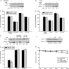
Protein kinase Cβ (PKCβ)‐mediated hepatocellular carcinoma cell motility and invasion depends on activation of extracellular signal‐regulating kinase (ERK)1/2–heat shock protein 27 (HSP27), and p38 mitogen‐activated protein kinase (MAPK)–HSP27. (a, b) HCCLM6 cells were pretreated with 4‐(4‐Fluorophenyl)‐2‐(4‐methylsulfinylphenyl)‐5‐(4‐pyridyl)1H‐imidazole (SB203580; 5 µM) for 2 h or 1.4‐diamino‐2.3‐dicyano‐1.4‐bis[2‐aminophenylthio] butadiene (U0126; 10 µM) for 1.5 h then stimulated by phorbol‐12‐myristate‐13‐acetate (PMA; 20 nM) incubation for 4 h. After 20 h, whole cell extracts from these samples were subjected to sodium dodecyl sulfate–polyacrylamide gel electrophoresis (SDS‐PAGE). Expression of total ERK1/2, p38 MAPK, and their phosphorylated forms were analyzed using specific antibodies by immunoblot analysis. The values underneath the bands represent the densitometric estimation of the relative density of the phosphorylated bands versus total protein bands comparing with that of HCCLM6 cells without treatment. (c,d) For observing the effects of SB203580 (c) and U0126 (d) on the migration and invasive ability of PMA‐stimulated HCCLM6 cells, in vitro migration and invasion assays were carried out in HCCLM6 cells, with the treatment outlined above, for 20 h in the presence of compounds described. The results represent means of three experiments. *P < 0.05 versus PMA‐treated HCCLM6 cells; **P < 0.01 versus HCCLM6 cells without treatment. (e) HCCLM6 cells were pretreated with U0126 (10 µM) or SB203580 (5 µM) followed by PMA (20 nM) for the indicated times. The blots were analyzed using a specific antibody against PKCβ and phosphor‐PKCβ. The values underneath the bands represent the densitometric estimation of the relative density of the phosphorylated bands comparing with that of HCCLM6 cells without treatment. (f) HCCLM6 cells were transfected with HSP27 small interfering RNA (siRNA) and negative control (non‐specific siRNA) for 48 h, equal amounts of cell extracts (30 µg) were subjected to SDS‐PAGE, the levels of total HSP27 was determined by immunoblot analysis. The level of glyceraldehyde‐3‐phosphate dehydrogenase (GAPDH) served as the loading control. The values underneath the bands represent the densitometric estimation of the relative density of the band, calculated by comparing the ratio of HSP27 versus GAPDH with that of HCCLM6 cells without siRNA treatment (the ratio in HCCLM6 cells without siRNA treatment was set as baseline 1.0). (g) After pretreatment with 100 nM HSP27 siRNA for 12 h, HCCLM6 cells were stimulated by PMA (20 nM) for 4 h then subjected to in vitro migration and invasion assays for 20 h. Non‐specific siRNA served as a control. The results represent means of three experiments. **P < 0.01 versus control. (h) HCCLM6 cells in a 96‐well plate were treated with 0.025 µM LY317615 or 20 nM PMA, 70 nM PKCβ siRNA, or 100 nM HSP27 for the indicated times. Cell survival was determined by 3‐(4,5‐dimethylthiazol‐2‐yl)‐2,5‐diphenyltetrazolium bromide assay. The results presented are means of three experiments; bars, ±SE.

Similar articles

Cited by

References

    1. Budhu AS, Zipser B, Forgues M, Ye QH, Sun Z, Wang XW. The molecular signature of metastases of human hepatocellular carcinoma. Oncology 2005; 69: 23–7. - PubMed
    1. Chambers AF, MacDonald IC, Schmidt EE, Morris VL, Groom AC. Clinical targets for anti‐metastasis therapy. Adv Cancer Res 2000; 79: 91–121. - PubMed
    1. Tang ZY. Hepatocellular carcinoma surgery – review of the past and prospects for the 21st century. J Surg Oncol 2005; 91: 95–6. - PubMed
    1. Christofori G. New signals from the invasive front. Nature 2006; 441: 444–50. - PubMed
    1. Wieser R. The transforming growth factor‐beta signaling pathway in tumorigenesis. Curr Opin Oncol 2001; 13: 70–7. - PubMed

Publication types

MeSH terms

Substances