Review
. 2008 Mar;99(3):459-66.
doi: 10.1111/j.1349-7006.2007.00704.x. Epub 2007 Dec 19.

Understanding tumor endothelial cell abnormalities to develop ideal anti-angiogenic therapies

Affiliations
Review

Understanding tumor endothelial cell abnormalities to develop ideal anti-angiogenic therapies

Kyoko Hida et al. Cancer Sci. 2008 Mar.

Abstract

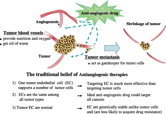
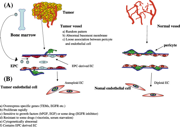
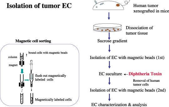
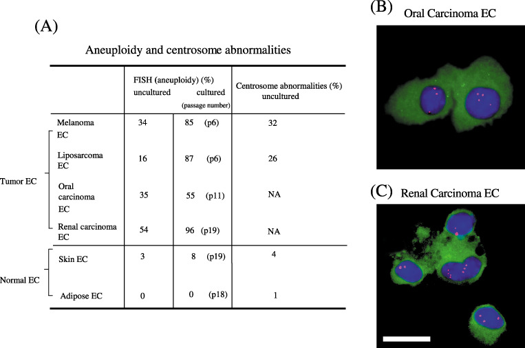
Tumor angiogenesis is necessary for solid tumor progression and metastasis. Tumor blood vessels have been shown to differ from their normal counterparts, for example, by changes in morphology. An important concept in tumor angiogenesis is that tumor endothelial cells are assumed to be genetically normal, even though these endothelial cells are structurally and functionally abnormal. To date, many anti-angiogenic drugs have been developed, but, their therapeutic efficacy is not dramatic and they have also been reported to cause toxic side effects. To develop ideal antiangiogenic therapies, understanding tumor endothelial cell abnormalities is important. We have isolated tumor endothelial cells from mouse tumor xenografts and have shown that tumor-associated endothelial cells are abnormal. Tumor-associated endothelial cells upregulate many genes, such as epidermal growth factor receptor (EGFR). Tumor-associated endothelial cells are also more sensitive to EGF. They also have relatively large, heterogeneous nuclei. Unexpectedly, tumor endothelial cells are cytogenetically abnormal. Fluorescence in situ hybridization (FISH) analysis showed that freshly isolated uncultured tumor endothelial cells were aneuploid and had abnormal multiple centrosomes. The degree of aneuploidy was exacerbated by passage in culture. In marked contrast, freshly isolated normal skin and adipose endothelial cells were diploid. They had normal centrosomes and remained cytogenetically stable in culture even up to 20 passages. We conclude that tumor endothelial cells can acquire cytogenetic abnormalities while in the tumor microenvironment. Questions as to whether or not tumor endothelial cells become resistant to antiangiogenic drugs are thus raised. Our preliminary data show that tumor endothelial cells are more resistant to certain chemotherapeutic drugs. Studies to evaluate the mechanism for cytogenetic abnormalities in tumor endothelial cells are underway. It is becoming quite clear that the tumor vasculature is much more complex and unpredictable than initially perceived. Here, we provide an overview of the current studies on tumor endothelial cell abnormalities.


Figures

Figure 1
Figure 1
Generally accepted concept of tumor angiogenesis and anti‐angiogenic therapy. Tumor blood vessels are important for tumor progression. They provide nutrition and oxygen to tumor tissue and get rid of waste. For tumor metastasis, tumor blood vessels are gatekeepers. Anti‐angiogenic therapy has been developed to target these blood vessels and inhibit new vascularization. Anti‐angiogenic therapy has been considered to have several advantages: (i) targeting endothelial cells might be a much more effective strategy than targeting tumor cells; (ii) tumor endothelial cells are the same among all tumor types, therefore an ideal anti‐angiogenic drug could be useful in treating all cancers; and (iii) tumor endothelial cells were, until recently, believed to be genetically stable. Thus, tumor endothelial cells might not acquire drug resistance, unlike tumor cells. However, recent studies suggest that tumor endothelial cells might be different from normal endothelial cells and might also be heterogeneous among organs or tumor types.
Figure 2
Figure 2
Differences in blood vessels and endothelial cells between tumors and normal tissues. (A) Tumor vessels are disorganized, whereas normal vasculature show a hierarchal branching pattern of arteries, veins, and capillaries. Tumor endothelial cell basement membranes have structural abnormalities including loose associations with endothelial cells, and various thicknesses of type IV collagen layers that are not usually seen in normal endothelial cells. Pericytes have abnormally loose associations with endothelial cells and extend cytoplasmic processes deep into the tumor tissue. Tumor vessels have chaotic blood flow and vessel leakiness due to loose endothelial cell interconnections. (B) Tumor endothelial cells differ from normal endothelial cells. (a) Tumor endothelial cells (EC) overexpress specific genes, such as tumor endothelial markers and epidermal growth factor receptors (EGFR); (b) tumor EC proliferate more rapidly and (c) are sensitive to growth factors such as basic fibroblast growth factor, EGF and vascular endothelial growth factor, or some drugs like EGFR inhibitors; (d) tumor EC are resistant to apoptotic stimuli such as serum starvation or chemotherapeutic drugs and (v) have cytogenetic abnormalities; (e) there are some endothelial progenitor cell‐derived endothelial cells in tumors.
Figure 3
Figure 3
Isolation of tumor endothelial cells. To isolate tumor endothelial cells (EC) from human tumor xenograft in nude mice, excised tissue is minced and digested with collagenase. After blood cells are removed by a single sucrose step‐gradient centrifugation, endothelial cells are isolated using a magnetic cell sorting (MACS) system using an anti‐CD31 antibody. Diphtheria toxin is added to tumor EC cultures to remove human tumor cells. After subculture for approximately 2 weeks, EC are purified by a second MACS using fluorescein‐isothiocyanate‐BS1‐B4 lectin. After EC purity is confirmed, the cells are characterized and used for analysis.
Figure 4
Figure 4
Cytogenetically abnormal tumor endothelial cells. (A) Quantitative analysis of cytogenetic abnormalities in endothelial cells. Tumor endothelial cells (melanoma, liposarcoma, oral carcinoma, and renal carcinoma endothelial cells) are aneuploid even before cultured and the degree of aneuploidy increases in culture, whereas uncultured normal endothelial cells are diploid and stay nearly diploid in culture. The centrosome abnormality was also detected in tumor endothelial cells. Mouse oral carcinoma endothelial cells (B) and renal carcinoma endothelial cells (C) were isolated and cytospun onto glass slides, followed by immunostaining with an anti‐CD31 antibody and fluorescent in situ hybridization with a chromosome 17 probe. Representative aneuploid endothelial cells are shown. Green, CD31; red; chromosome 17; blue, 4′,6′‐diamidino‐2‐phenylindole dihydrochloride. Scale bar, 10 µm.
Figure 5
Figure 5
Possible mechanisms causing tumor endothelial cytogenetic abnormalities include the following. (a) Tumor cell transdifferentiation. In the case of hematopoietic tumors, a common progenitor targeted by transformation can differentiate in tumor cells or endothelial cells. (b) Dedifferentiation. Tumor cells might also dedifferentiate to endothelial cells. (c) Tumor microenvironment. Factors such as growth factors or cytokines in the tumor microenvironment could cause genetic instability. Hypoxia in tumors is known to cause genetic changes, for example, upregulation of survival factors, not only in tumor cells. Thus, the tumor microenvironment can induce genetic instability not only tumor cells, but also endothelial cells. (d) Cell fusion. Malignant tumor cells can fuse with normal endothelial cells or circulating endothelial progenitor cells (EPC). (e) Uptake of oncogene or gene transfer. Endothelial cells can uptake human tumor oncogenes released from EPC or tumor cells by phagocytosis of apoptotic bodies or microvesicles (MV).

References

    1. Folkman J. Tumor angiogenesis: therapeutic implications. N Engl J Med 1971; 285: 1182–6. - PubMed
    1. Folkman J, Kerbel R. Role of angiogenesis in tumor growth and metastasis Clinical translation of angiogenesis inhibitors. Semin Oncol 2002; 29: 15–18. - PubMed
    1. Folkman J. Angiogenesis: an organizing principle for drug discovery? Nat Rev Drug Discov 2007; 6: 273–86. - PubMed
    1. Johnson DH, Fehrenbacher L, Novotny WF et al . Randomized phase II trial comparing bevacizumab plus carboplatin and paclitaxel with carboplatin and paclitaxel alone in previously untreated locally advanced or metastatic non‐small‐cell lung cancer. J Clin Oncol 2004; 22: 2184–91. - PubMed
    1. Keedy VL, Sandler AB. Inhibition of angiogenesis in the treatment of non‐small cell lung cancer. Cancer Sci 2007; 98: 1825–30. - PMC - PubMed

Publication types

Substances