. 2008 Jan 2;3(1):e1400.
doi: 10.1371/journal.pone.0001400.

Variability and action mechanism of a family of anticomplement proteins in Ixodes ricinus

Affiliations

Variability and action mechanism of a family of anticomplement proteins in Ixodes ricinus

Bernard Couvreur et al. PLoS One. .

Abstract

Background: Ticks are blood feeding arachnids that characteristically take a long blood meal. They must therefore counteract host defence mechanisms such as hemostasis, inflammation and the immune response. This is achieved by expressing batteries of salivary proteins coded by multigene families.

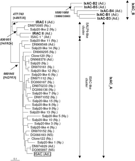
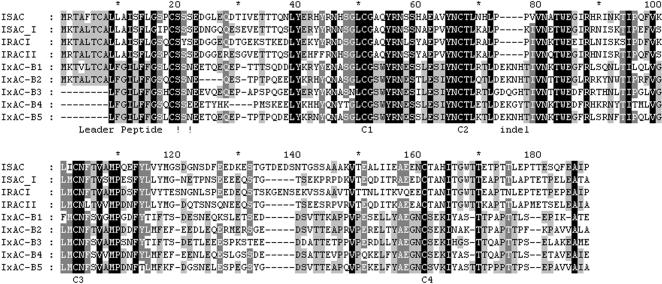
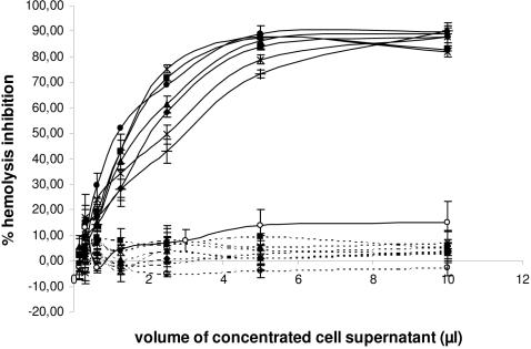
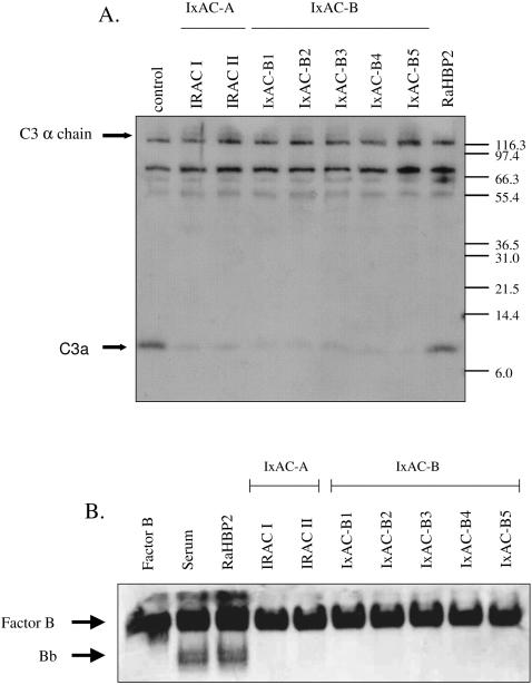
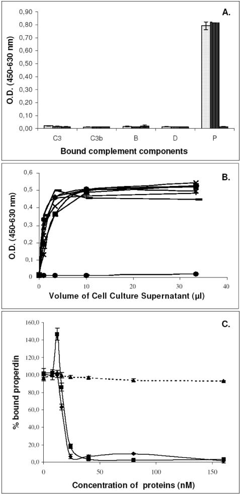
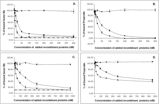
Methodology/principal findings: We report the in-depth analysis of a tick multigene family and describe five new anticomplement proteins in Ixodes ricinus. Compared to previously described Ixodes anticomplement proteins, these segregated into a new phylogenetic group or subfamily. These proteins have a novel action mechanism as they specifically bind to properdin, leading to the inhibition of C3 convertase and the alternative complement pathway. An excess of non-synonymous over synonymous changes indicated that coding sequences had undergone diversifying selection. Diversification was not associated with structural, biochemical or functional diversity, adaptation to host species or stage specificity but rather to differences in antigenicity.

Conclusions/significance: Anticomplement proteins from I. ricinus are the first inhibitors that specifically target a positive regulator of complement, properdin. They may provide new tools for the investigation of role of properdin in physiological and pathophysiological mechanisms. They may also be useful in disorders affecting the alternative complement pathway. Looking for and detecting the different selection pressures involved will help in understanding the evolution of multigene families and hematophagy in arthropods.


Conflict of interest statement

Competing Interests: The activities of IxAC-B1 to B5 are being patented.

Figures

Figure 1
Figure 1. Phylogenetic analysis of Ixodes anticomplement proteins.
A distance dendrogram was constructed from an alignment of 41 tick mature anticomplement proteins using programs in the Phylip 3.65 package (see text). Branch length is proportional to distances between peptide sequences. The bootstrap values are indicated near major nodes, calculated from 1000 replicates of the peptide and nucleotide sequence alignments, respectively. Bold characters: I. ricinus entries; *: I. pacificus sequence; all others are from I. scapularis. Prototypical ISAC is boxed. Sequences are identified by their accession number in databases or by descriptive names when available. Ad., isolated from adults; Ny., isolated from nymphs; NS, not specified.
Figure 2
Figure 2. Alignment of Ixodes anticomplement proteins.
The 7 anticomplement proteins from I. ricinus (IRAC I and II; IxAC-B1 to B5) were aligned with the prototypical anticomplement protein from I. scapularis (ISAC) and the homolog from I. pacificus (ISAC I). Individual residues printed in white on a black background are conserved in all 9 aligned sequences; white residues on a grey background are conserved in 7 or 8 of 9 entries; black residues on a grey background are conserved in 5 or 6 entries; black residues on a white background are conserved in less than 5 entries. -, gap; ! !, region of predicted signal peptide cleavage; C1 to C4, conserved cysteine residues.
Figure 3
Figure 3. Western blot analysis of recombinant IxAC-V5His proteins from I. ricinus.
Standardised amounts of recombinant IxAC-V5His proteins from supernatants of transfected 293T cells were analysed by SDS/PAGE and detected by western blotting using an anti-V5 monoclonal antibody. A) Parallel analysis of IxACs, B) N-deglycosylation of IxAC-B1-V5His.1, untreated, 2, incubated with PNGase (New England Biolabs).
Figure 4
Figure 4. Comparison of Ixodes anticomplement protein tertiary structure.
Aligned IxAC amino-acid sequences from I. ricinus were submitted to hydrophobic cluster analysis (HCA). Groups of adjacent hydrophobic residues are outlined and shaded. Proline (asterisk), glycine (open rectangle), serine (dotted square) and threonine (open square) are highlighted. The overall distribution of hydrophobic clusters and their size, shape and orientation are very similar.
Figure 5
Figure 5. Effect of recombinant I. ricinus IxAC proteins on the alternative and classical pathways of complement activation.
Assays of the alternative (AP, solid lines) and classical (CP, dashed lines) complement activation pathway were conducted in the presence of normalized amounts of recombinant I. ricinus IxACs produced in the supernatant of transfected 293T cells. Values for the percent inhibition of rabbit red blood cell lysis in the presence of human serum are indicated. The values are means ± standard deviation of triplicates. RaHBP2 was used as negative control. Black diamond :IRAC I; black square:IRAC II; black triangle:IxAC-B1; cross:IxAC-B2; star:IxAC-B3; closed circle:IxAC-B4; Plus:IxAC-B5; open circle:RaHBP2.
Figure 6
Figure 6. Inhibition of C3a formation and factor B cleavage.
Aliquots of supernatant from AP hemolysis assays conducted in the presence of standardized amounts of IxACs from I. ricinus and unrelated control RaHBP2 were analyzed by western blotting. Panel A: Blots from gels run under denaturing conditions were probed with monospecific anti-C3a serum. The α-chain of precursor C3 (116 kDa) and the C3a peptide (∼10 kDa) are indicated by arrows. Panel B: Blots from gels run under non-denaturing conditions were probed with a antiserum to factor B. Purified factor B was used as a positive control.
Figure 7
Figure 7. ELISA analysis of the binding of IxAC proteins to immobilized C3 convertase components.
Panel A: Binding of IxACs to AP components. Purified recombinant IRAC II, IxAC-B1 or unrelated protein Iris were added to microtiter wells previously coated with purified factors C3, C3b, fB, fD or properdin (P). Bound proteins were detected with an anti-V5 monoclonal antibody using an ELISA format. Light dotted histogram: IRAC II; dark dotted histogram: IxAC-B1; black histogram: Iris. Panel B: Increasing amounts of normalized supernatant from transfected culture 293T cells were added to immobilized properdin. Bound IxACs were detected with an anti-V5 antibody. Black diamond: Iris; black square: IRAC I; black triangle: IRAC II; cross: IxAC-B1; star: IxAC-B2; closed circle: IxAC-B3; plus: IxAC-B4; minus : IxAC-B5. Panel C. Competition between properdin and IxACs for C3b binding. Purified properdin and increasing amounts of IRAC II, IxAC-B1 or unrelated control IRIS were added simultaneously to C3b-precoated microtiter wells. Bound properdin was detected with an anti-properdin monoclonal antibody. Black diamond: IRAC II; black square: IxAC-B1; black triangle: Iris.
Figure 8
Figure 8. Effect of IxAC proteins on the formation and stability of C3 convertase.
Panel A and B: The effect of IxAC proteins on the formation of C3 convertase was evaluated by incubating simultaneously purified factors B, D and properdin with increasing amounts of IRAC II or IxAC-B1 on C3b-coated wells. Panel C and D: The effect of IxAC proteins on the stability of C3 convertase was assessed by incubating preformed C3 convertase (fB, fD and properdin pre-incubated for 1 hour) on C3b-coated plates with increasing amounts of recombinant IxACs. Bound factor B or properdin was detected with an anti-factor B antibody (A–C) or anti-properdin antibody (B–D), respectively. Recombinant IRIS was used as negative control. Black diamond: IRAC II; black square: IxAC-B1; black triangle: IRIS.
Figure 9
Figure 9. Effect of IxAC proteins on the deposition of C3b and factor B on agarose-coated plates.
Loading human serum on agarose-coated microplate wells activates the alternative complement pathway. Purified recombinant IRAC II (A–B) or IxAC-B1 (C–D) were added after 30, 45 or 60 minutes. The reactions were stopped at various times. C3b and factor B deposition was detected using anti-C3 antibody (A–C) or anti-factor B antibody (B–D), respectively. Black diamond: no added protein; black square: 30 min.; black triangle: 45 min.; cross: 60 min.
Figure 10
Figure 10. Expression patterns of individual IxACs.
PolyA+ RNA was extracted from various I. ricinus material as indicated and reverse transcribed. The resulting cDNAs were submitted to PCR analysis using pairs of primers specific for the indicated IxACs. Non- reverse transcribed polyA+ RNA from the pool was included as negative control (Ctrl). PCR products were run on 1.2 % agarose gels. M, DNA size markers. B1 to B5, IxAC-B1 to IxAC-B5. Sizes in bp are indicated. Panel A: Analysis of salivary glands of a tick population (70 specimens, pool) and from individual female ticks at day 5 of the bloodmeal (A to J). Panel B: Analysis of pooled salivary glands of tick female populations at day 0 (25 specimens), day 3 (25 specimens) and day 5 (70 specimens) of the bloodmeal as well as from pooled gorged nymphs (25 specimens) and larvae (25 specimens).
Figure 11
Figure 11. Antigenic specificity of recombinant IxACs from I. ricinus.
Standardised amounts of the seven I. ricinus recombinant IxACs were analysed for antigenic specificity. Panel A. Western blot analysis. The serum from a mouse immunized against IxAC-B1 by genetic immunization followed by a protein boost recognised solely recombinant IxAC B1. M, molecular weight markers (Mark12, Invitrogen). Panel B. Seroneutralization experiments. AP hemolysis assays were conducted with and without the seven recombinant IxACs. 100% hemolysis was obtained in the absence of anticomplement protein (light dotted histogram). Recombinant IxACs alone (dark dotted histogram) or recombinant IxACs plus heat-inactivated sera from mice immunized against IxAC-B1 (anti IxAC-B1, black histogram) or mock immunized mice (anti-PBS, white histogram) were added as indicated. Neutralization of activity as indicated by a recovery of hemolysis, was observed only on IxAC-B1. Upper panels: seroneutralization of IxAC-A subfamily. Lower panel: seroneutralization of IxAC-B subfamily. Error bars represent standard deviations.

References

    1. Price PW. Princeton, New Jersey: Princeton University Press; 1980. Evolutionary evolutionary biology of parasites. Monographs in population biology. p. 237. - PubMed
    1. Jongejan F, Uilenberg G. The global importance of ticks. Parasitology. 2004;129:S3–S14. - PubMed
    1. Sonenshine DE. New York, Oxford: Oxford University Press; 1991. Biology of ticks. Vol. I.
    1. Mans BJ, Gothe R, Neitz AWH. Biochemical perspectives on paralysis and other forms of toxicoses caused by ticks. Parasitology. 2004;129:S95–S111. - PubMed
    1. Ribeiro JMC. Role of saliva in blood-feeding by arthropods. Ann Rev Entomol. 1987a;32:463–478. - PubMed

Publication types