. 2008 Jan 29;47(4):1144-56.
doi: 10.1021/bi701807c. Epub 2008 Jan 3.

Outer segment oligomerization of Rds: evidence from mouse models and subcellular fractionation

Affiliations

Outer segment oligomerization of Rds: evidence from mouse models and subcellular fractionation

Dibyendu Chakraborty et al. Biochemistry. .

Abstract

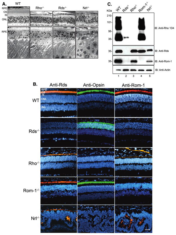
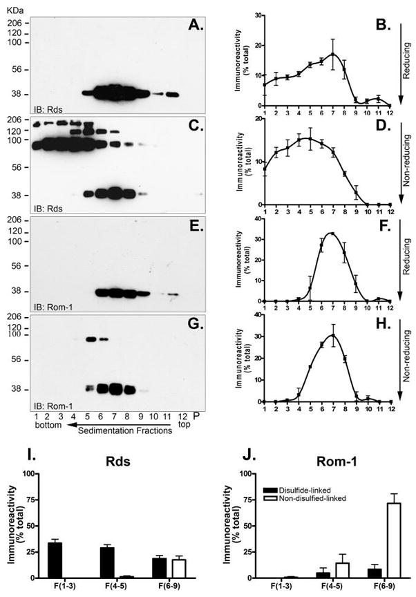
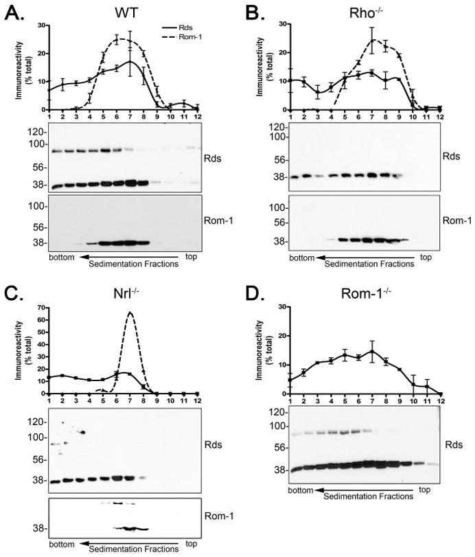
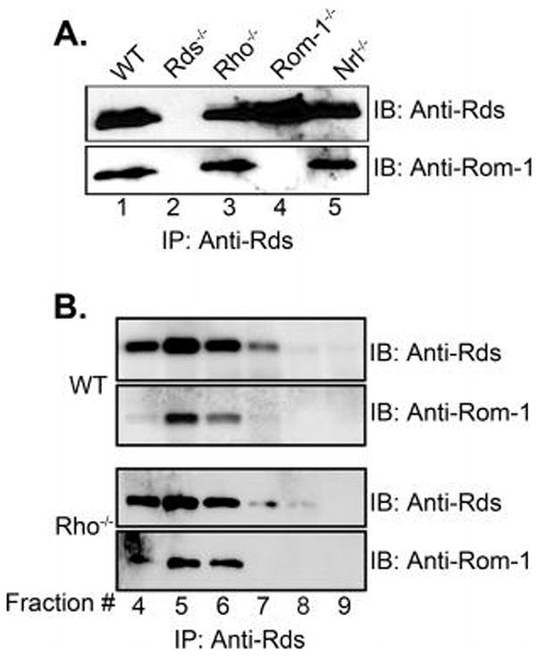
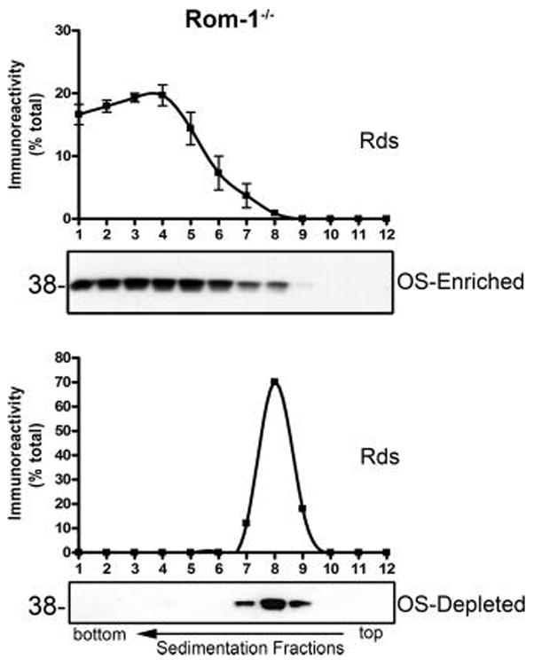
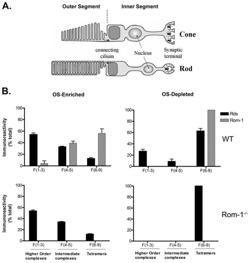
Retinal degeneration slow (Rds) is a photoreceptor-specific tetraspanin glycoprotein essential for photoreceptor outer segment (OS) morphogenesis. Over 80 mutations in this protein are associated with several different retinal diseases. Rds forms a mixture of disulfide-linked homomeric dimers, octamers, and higher-order oligomers, with Cys150 playing a crucial role in its oligomerization. Rds also forms noncovalent homo- and hetero-tetramers with its nonglycosylated homologue, Rom-1. Here, we evaluated the subcellular site of Rds oligomerization and the pattern of Rds/Rom-1 complex assembly in several types of knockout mice, including rhodopsin (Rho-/-, lacking rod OS), Rom-1 (Rom-1-/-), neural retina leucine zipper (Nrl-/-, cone-dominant), and in comparison with wild-type (WT, rod-dominant) mice. Oligomerization and the pattern of complex assembly were also evaluated in OS-enriched vs OS-depleted preparations from WT and Rom-1-/- retinas. Velocity sedimentation under reducing- and nonreducing conditions and co-immunoprecipitation experiments showed the presence of Rds mainly as homo- and hetero-tetramers with Rom-1 in the photoreceptor inner segment (IS), while higher-order, disulfide-linked intermediate complexes and oligomers were exclusively present in the photoreceptor OS. Rom-1-independent oligomerization of Rds was observed in Rom-1-/- retinas. The pattern of Rds complexes in cones from Nrl-/- mice was comparable to that in rods from WT mice. On the basis of these findings, we propose that Rds traffics from the IS to the OS as homo- and hetero-tetramers, with subsequent disulfide-linked oligomerization occurring concomitant with OS disc morphogenesis (at either the base of OS or the tip of the connecting cilium). These results suggest that Rds mutations that interfere with tetramer formation can block Rds trafficking to the OS, leading to loss-of-function defects.


Figures

Figure 1
Figure 1
Retinal morphology, expression levels and cellular localization of Rds and Rom-1 from mouse retinas. (A) Upper panels: LM images of retinas from 1-month old WT, Rho−/−, Rds−/−, and Nrl−/− mice. Lower panels: corresponding EM images of photoreceptor-RPE interface from these animals. Scale bar for LM and EM images are 25 and 4 μM, respectively. EM demonstrates lack of OSs in Rho−/− and Rds−/− retinas. (B) Immunohistochemistry of retinas from 1-month old WT, Rds−/−, Rho−/−, Rom-1−/−, and Nrl−/− mice. Left-hand panels; anti-Rds immunoreactivity; middle panels: anti-rhodopsin; right-hand panels; anti-Rom-1. Nuclei were stained with DAPI (blue). Red and green fluorescence is due to Cy3 and FITC conjugated secondary antibodies. Rho−/− retinas exhibit immunolabeling for Rds and Rom-1 at the tip of the connecting cilium. Abbreviations: RPE, retinal pigment epithelium; OS, outer segment; IS, inner segment; ONL, outer nuclear layer; INL, inner nuclear layer. (C) Western blot analysis of WT, Rds−/−, Rho−/−, Rom-1−/−, and Nrl−/− mouse retinas at p30, probed with antibodies against rhodopsin, Rds, Rom-1, and actin (control). Note marked reduction of anti-rhodopsin immunostaining in the Rds−/− retina (photoreceptor dysplasia, absence of OSs) and coordinate loss of Rom-1 expression. In Nrl−/− retina, note lack of rhodopsin expression (lacks rod photoreceptors) and also reduced Rom-1/Rds ratio compared to WT and Rho−/− retinas. Numerical values in the left-hand margin (Mr, in kDa) of each panel indicate the migration positions of protein molecular weight markers.
Figure 2
Figure 2
Subunit assembly of Rds and Rom-1 in the rod-dominant WT retina. Whole WT retinas (200 μg of protein) were solubilized in the presence of either DTT (reducing conditions, panels A and E) or NEM (nonreducing conditions, panels C and G). Extracts were sedimented under reducing (panels A and E) or nonreducing (panels C and G) conditions. Western blot analysis was performed on all fractions and probed with anti-Rds (panels A and C and corresponding quantification in panels B and D) and anti-Rom-1 (panels E and G and corresponding quantification in panels F and H). Levels of Rds (panel I) and Rom-1 (panel J) present in noncovalently linked vs disulfide-linked complexes were averaged from three independent experiments (results presented as % of the total). Under reducing conditions, a single peak with tetrameric stoichiometry was observed for both proteins, spanning fractions 5 to 10. Under nonreducing conditions, higher-order Rds homo-oligomers linked through disulfide bonds were detected in fractions 1–4. By contrast, Rom-1 mainly appeared as one main peak corresponding to tetrameric complexes, with only a small portion representing disulfide-linked complexes detected in fractions 5 and 6. Numerical values in the left-hand margin (Mr, in kDa) of each panel indicate the migration positions of protein molecular weight markers.
Figure 3
Figure 3
Subunit assembly of Rds and Rom-1 in retinas from knockout mice. Whole retina extracts (200 μg of protein each) from WT (A), Rho−/− (B), Nrl−/− (C), and Rom-1−/− (D) mice were sedimented under nonreducing conditions. Identical Western blots under reducing conditions of the fractions from each extract were probed with anti-Rds and anti-Rom-1 antibodies. Rds homo-oligomeric complexes are much less abundant in the Rho−/− retina compared to WT, and the majority of Rds and Rom-1 forms homo- and hetero-tetrameric complexes. In contrast, Nrl−/− and Rom-1−/− retinas show WT-like sedimentation patterns with respect to Rds distribution and oligomerization. Numerical values in the left-hand margin (Mr, in kDa) of each panel indicate the migration positions of protein molecular weight markers.
Figure 4
Figure 4
Association of Rds and Rom-1 in retinas from different mouse strains. (A) Immunoprecipitation of retinal extracts from all models used in this study with anti-Rds, and corresponding Western blots probed with anti-Rds (upper panel) and anti-Rom-1 (lower panel) antibodies. Rds and Rom-1 interact in IS-enriched Rho−/− (OS-free) and Nrl−/− (pure cone, rod-free) retinas. Lanes are numbered 1–5 to reflect the genotype of the extract loaded in each lane. (B) Western blot analysis of immunoprecipitates of fractions 4–9 from WT and Rho−/− retinas. Blots were probed with anti-Rds and anti-Rom-1. Fractions 5 and 6 represent mostly Rds/Rom-1 hetero-tetramers. Lanes are numbered 4–9 to reflect the fraction number obtained from the sucrose gradient sedimentation.
Figure 5
Figure 5
Oligomerization of Rds in the outer segment. (A) Reducing velocity sedimentation analysis of OS-enriched and OS-depleted preparations from WT retinas and retinal extracts from Rho−/− mice. Immunoblots were probed with anti-Rds and anti-Rom-1 antibodies. In the presence of DTT, higher-order complexes of Rds and Rom-1 were reduced to tetramers that appeared as a single peak. (B) Nonreducing velocity sedimentation of OS-enriched and OS-depleted preparations from WT retinas. Note mainly the presence of higher-order complexes of Rds in the OS-enriched preparation, whereas the OS-depleted samples show mainly the presence of tetrameric forms. The migration position of Rds and Rom-1 monomers (Mr ~ 38 kDa) is indicated.
Figure 6
Figure 6
Rds complex assembly in the absence of Rom-1. Velocity sedimentation analysis of OS-enriched and OS-depleted fractions from Rom-1−/− retinas performed under nonreducing conditions and immunobloted with anti-Rds antibody. The majority of Rds is present in octamers and higher-order homo-oligomers (fractions 1–6), similar to findings with WT OS preparations. Note the complete lack of Rds higher-order oligomers in the OS-depleted samples from Rom-1−/− retina, which contain mainly Rds homo-tetramers (fraction 8). This finding suggests that Rds tetrameric form is the preferred complex that traffics Rds from the IS to the OS, Rds homo-oligomers are essential for OS rim structure formation, and that Rds higher-order oligomerization occurs in the OS. The migration position of Rds (Mr ~ 38 kDa) is indicated.
Figure 7
Figure 7
Summary illustration depicting the sites of Rds and Rom-1 complex formation in rod and cone photoreceptor cells. (A) Structural diagram of rod and cone photoreceptor cell. (B) Distribution of Rds complexes in OS-enriched and OS-depleted preparations from WT and Rom-1−/− retinas. The histograms summarize the results of the present study. Depending on the nature of the complex, Rds was recovered mostly as covalent disulfide-linked high order oligomers in the OS and noncovalent tetrameric complex in the IS from both models. Some of these complexes are homo complexes of Rds or hetero associations with Rom-1. The rearrangement from a noncovalent tetrameric complexes formed in the IS to a covalent octameric and oligomeric complexes formed in the OS would require the enzymatic action of an as yet unknown disulfide isomerase that should be located in the OS.

References

    1. Molday RS, Hicks D, Molday L. Peripherin. A rim-specific membrane protein of rod outer segment discs. Invest Ophthalmol Vis Sci. 1987;28:50–61. - PubMed
    1. Bascom RA, Manara S, Collins L, Molday RS, Kalnins VI, McInnes RR. Cloning of the cDNA for a novel photoreceptor membrane protein (rom-1) identifies a disk rim protein family implicated in human retinopathies. Neuron. 1992;8:1171–1184. - PubMed
    1. Sanyal S, Jansen HG. Absence of receptor outer segments in the retina of Rds mutant mice. Neurosci Lett. 1981;21:23–26. - PubMed
    1. Travis GH, Sutcliffe JG, Bok D. The retinal degeneration slow (Rds) gene product is a photoreceptor disc membrane-associated glycoprotein. Neuron. 1991;6:61–70. - PubMed
    1. Kajiwara K, Hahn LB, Mukai S, Travis GH, Berson EL, Dryja TP. Mutations in the human retinal degeneration slow gene in autosomal dominant retinitis pigmentosa. Nature. 1991;354:480–483. - PubMed

Publication types

MeSH terms

LinkOut - more resources