. 2008 Jan 2:9:1.
doi: 10.1186/1471-2202-9-1.

Amiloride-sensitive channels in type I fungiform taste cells in mouse

Affiliations

Amiloride-sensitive channels in type I fungiform taste cells in mouse

Aurelie Vandenbeuch et al. BMC Neurosci. .

Abstract

Background: Taste buds are the sensory organs of taste perception. Three types of taste cells have been described. Type I cells have voltage-gated outward currents, but lack voltage-gated inward currents. These cells have been presumed to play only a support role in the taste bud. Type II cells have voltage-gated Na+ and K+ current, and the receptors and transduction machinery for bitter, sweet, and umami taste stimuli. Type III cells have voltage-gated Na+, K+, and Ca2+ currents, and make prominent synapses with afferent nerve fibers. Na+ salt transduction in part involves amiloride-sensitive epithelial sodium channels (ENaCs). In rodents, these channels are located in taste cells of fungiform papillae on the anterior part of the tongue innervated by the chorda tympani nerve. However, the taste cell type that expresses ENaCs is not known. This study used whole cell recordings of single fungiform taste cells of transgenic mice expressing GFP in Type II taste cells to identify the taste cells responding to amiloride. We also used immunocytochemistry to further define and compare cell types in fungiform and circumvallate taste buds of these mice.

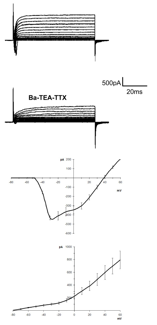
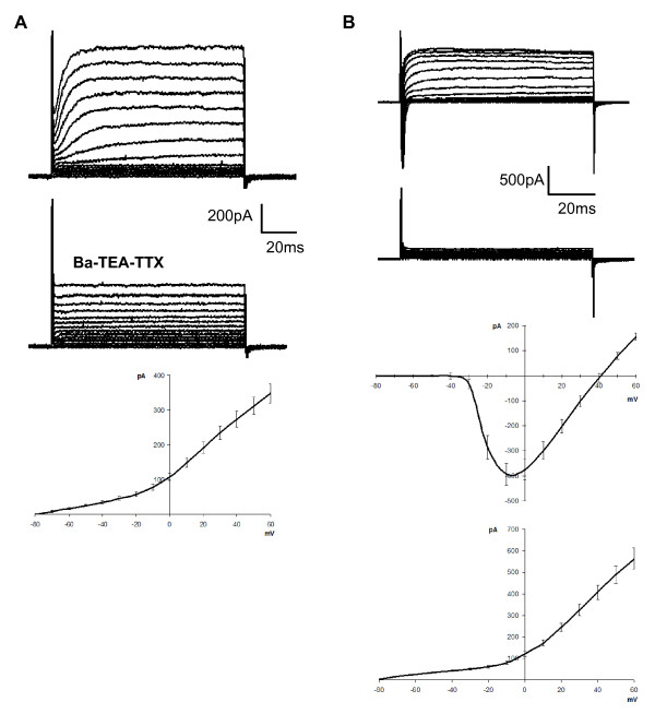
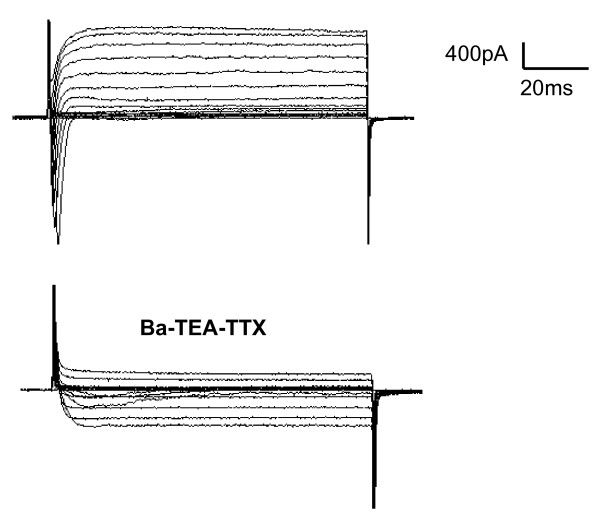
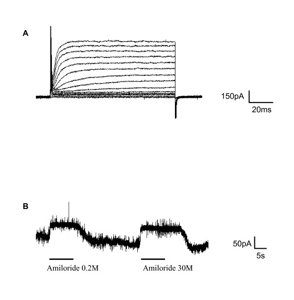
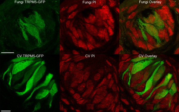
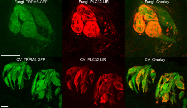
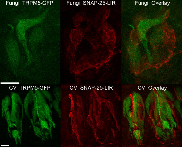
Results: Taste cell types were identified by their response to depolarizing voltage steps and their presence or absence of GFP fluorescence. TRPM5-GFP taste cells expressed large voltage-gated Na+ and K+ currents, but lacked voltage-gated Ca2+ currents, as expected from previous studies. Approximately half of the unlabeled cells had similar membrane properties, suggesting they comprise a separate population of Type II cells. The other half expressed voltage-gated outward currents only, typical of Type I cells. A single taste cell had voltage-gated Ca2+ current characteristic of Type III cells. Responses to amiloride occurred only in cells that lacked voltage-gated inward currents. Immunocytochemistry showed that fungiform taste buds have significantly fewer Type II cells expressing PLC signalling components, and significantly fewer Type III cells than circumvallate taste buds.

Conclusion: The principal finding is that amiloride-sensitive Na+ channels appear to be expressed in cells that lack voltage-gated inward currents, likely the Type I taste cells. These cells were previously assumed to provide only a support function in the taste bud.


Figures

Figure 1
Figure 1
Whole-cell voltage-gated currents in isolated TRPM5-GFP-positive fungiform taste cells. In GFP-labeled cells expressing TRPM5 channels, depolarizing steps from -80 mV elicit voltage-gated inward and outward currents. The outward current is mostly blocked by TEA indicating the involvement of voltage-gated K+ channels while the inward current was blocked by TTX indicating the involvement of voltage-gated Na+ channels. Replacement of Ca2+ with Ba2+ did not reveal an inward current suggesting that these cells do not express voltage-gated Ca2+ channels. The I-V plot is represented for both inward and outward currents in Tyrodes (n = 6 cells; mean ± sem).
Figure 2
Figure 2
Whole-cell voltage-gated currents in unlabeled fungiform taste cells of the TRPM5-GFP mice. A: Some unlabeled cells (53%) exhibit only an outward current in response to depolarizing voltage steps. The Barium-TEA-TTX solution decreased the outward current indicating the involvement of voltage-dependent K+ channels. The I-V plot is represented (n = 52 cells; mean ± sem). B: Other cells (46%) exhibit outward K+ and inward Na+ currents, but lack a voltage-dependent Ca2+ (Ba2+) current. The I-V plot is represented for inward and outward currents (n = 45 cells; mean ± sem).
Figure 3
Figure 3
Whole-cell voltage-gated currents in an unlabeled fungiform taste cell expressing a Ca2+ current. In one cell, showing a large inward Na+ and outward current K+ in response to depolarizing voltage steps, the application in of the Barium-TEA-TTX solution elicited a Ca2+ current, typical of type III cells.
Figure 4
Figure 4
Amiloride effect on the steady-state current in a fungiform taste cell. A: Cells exhibiting an amiloride effect respond to depolarizing voltage steps with only outward currents. B: The application of amiloride in the bath, at 30 or 0.2 μM, decreased the steady-state current (holding potential -100 mV).
Figure 5
Figure 5
Expression of TRPM5-GFP and Propidium Iodide in confocal images of fungiform and circumvallate taste buds. Propidium Iodide was used to stain the nucleus of all types of cells (red) in a taste bud. Using the same section thickness, fungiform taste buds (top figures) and circumvallate taste buds (bottom figures) showed the same number of cells. Scale bar: 10 μm.
Figure 6
Figure 6
Expression of TRPM5-GFP and PLCβ2 in confocal images of fungiform and circumvallate taste buds. The top figures illustrate the expression of GFP under the control of the TRPM5 promoter (green) and PLCβ2-ir (red) in fungiform taste buds. The bottom figures illustrate labeling in circumvallate taste buds. Note that in circumvallate taste buds there are far more TRPM5-GFP cells relative to fungiform taste buds, however PLCβ2 and TRPM5-GFP were generally co-localized in both fungiform and circumvallate taste buds. Each figure represents merged images from a Z-series. Scale bar: 10 μm.
Figure 7
Figure 7
Expression of TRPM5-GFP and SNAP-25 in confocal images of fungiform and circumvallate taste buds. The top figures illustrate the expression of GFP under the control of the TRPM5 promoter (green) and SNAP-25-ir (red) in fungiform taste buds and the bottom figures illustrate labeling in circumvallate taste buds. No co-localization was observed between TRPM5-GFP and SNAP-25-ir. The number of SNAP-25-ir and GFP-labeled cells was larger in circumvallate than in fungiform taste buds. Each figure represents merged images from a Z-series. Scale bar: 10 μm.

Similar articles

Cited by

References

    1. Finger TE., Simon, S.A. Cell Biology of Taste Epithelium. In Neurobiology of Taste and Smell, 2nd edition Edited by Finger, TE, Silver, WL, Restrepo, D New York: Wiley Liss. 2000. pp. 287–314.
    1. Kinnamon SC., Margolskee, R.F. Taste Transduction. In Handbook of Senses Volume 1 Edited by Firestein, S, Smith, D, Shepherd, G In Press. 2007.
    1. Pumplin DW, Yu C, Smith DV. Light and dark cells of rat vallate taste buds are morphologically distinct cell types. J Comp Neurol. 1997;378:389–410. doi: 10.1002/(SICI)1096-9861(19970217)378:3<389::AID-CNE7>3.0.CO;2-#. - DOI - PubMed
    1. Lawton DM, Furness DN, Lindemann B, Hackney CM. Localization of the glutamate-aspartate transporter, GLAST, in rat taste buds. Eur J Neurosci. 2000;12:3163–3171. doi: 10.1046/j.1460-9568.2000.00207.x. - DOI - PubMed
    1. Bartel DL, Sullivan SL, Lavoie EG, Sevigny J, Finger TE. Nucleoside triphosphate diphosphohydrolase-2 is the ecto-ATPase of type I cells in taste buds. J Comp Neurol. 2006;497:1–12. doi: 10.1002/cne.20954. - DOI - PMC - PubMed

Publication types

MeSH terms

LinkOut - more resources