. 2008 Apr;162(1):94-107.
doi: 10.1016/j.jsb.2007.11.009. Epub 2007 Nov 29.

Structures of open (R) and close (T) states of prephenate dehydratase (PDT)--implication of allosteric regulation by L-phenylalanine

Affiliations

Structures of open (R) and close (T) states of prephenate dehydratase (PDT)--implication of allosteric regulation by L-phenylalanine

Kemin Tan et al. J Struct Biol. 2008 Apr.

Abstract

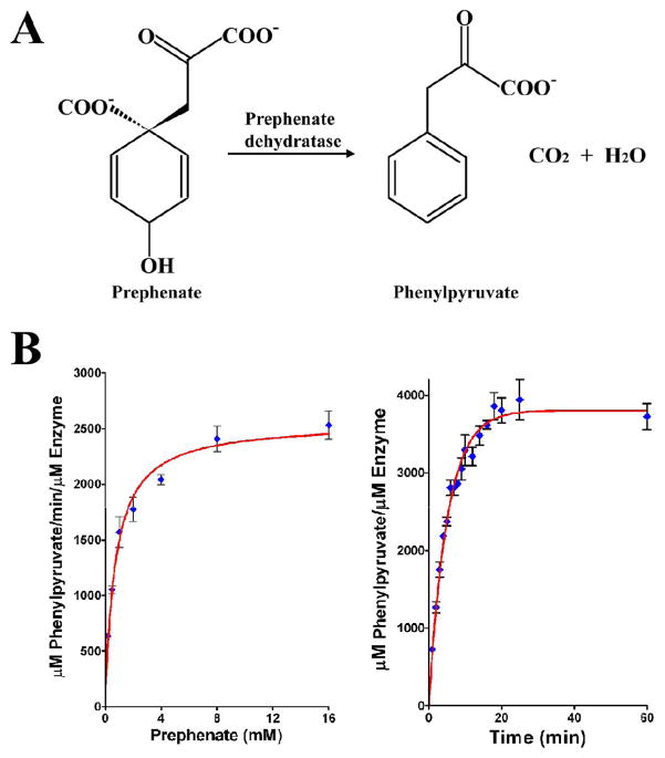
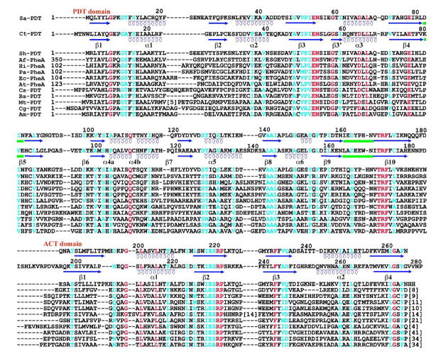
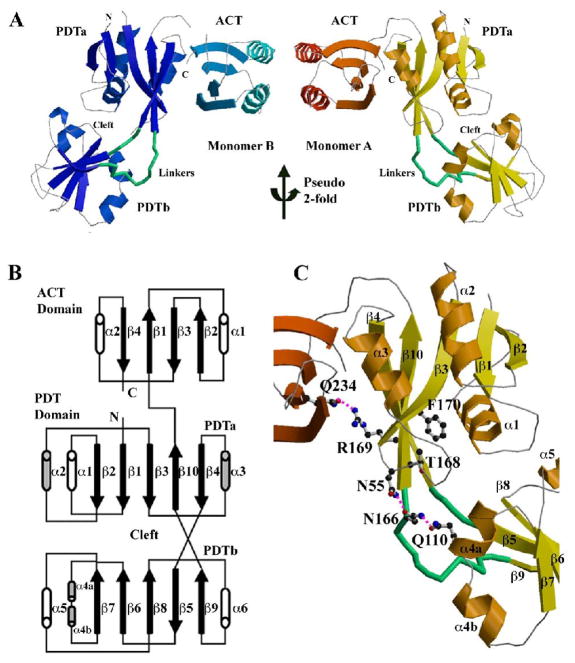
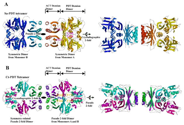
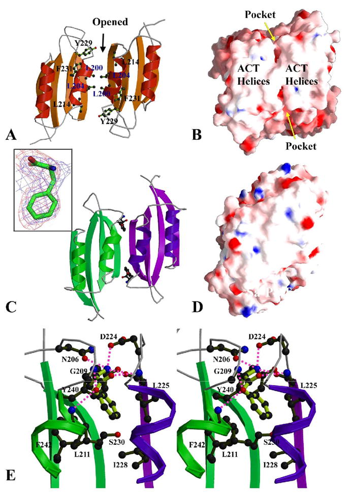
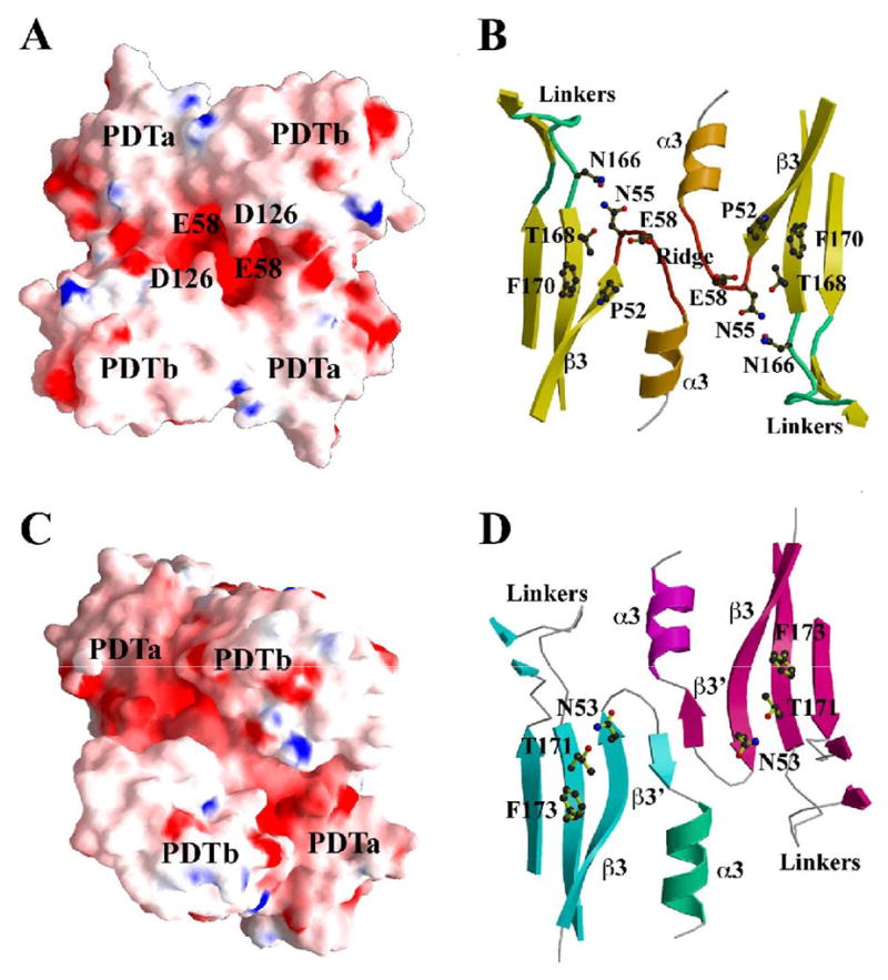
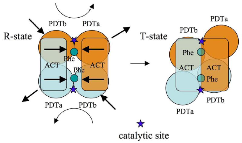
The enzyme prephenate dehydratase (PDT) converts prephenate to phenylpyruvate in L-phenylalanine biosynthesis. PDT is allosterically regulated by L-Phe and other amino acids. We report the first crystal structures of PDT from Staphylococcus aureus in a relaxed (R) state and PDT from Chlorobium tepidum in a tense (T) state. The two enzymes show low sequence identity (27.3%) but the same prototypic architecture and domain organization. Both enzymes are tetramers (dimer of dimers) in crystal and solution while a PDT dimer can be regarded as a basic catalytic unit. The N-terminal PDT domain consists of two similar subdomains with a cleft in between, which hosts the highly conserved active site. In one PDT dimer two clefts are aligned to form an extended active site across the dimer interface. Similarly at the interface two ACT regulatory domains create two highly conserved pockets. Upon binding of the L-Phe inside the pockets, PDT transits from an open to a closed conformation.


Figures

Figure 1
Figure 1
The pathway and the enzymatic activity of Sa-PDT. (A) The conversion of prephenate to phenylpyruvate by prephenate dehydratase (PDT). (B) Activity assay for PDT was performed as described by Dopheide et al. (28) and as described in Materials and Methods. The left panel shows the reaction rate at different prephenate concentrations, the enzyme was 0.04 μM and the prephenate concentrations vary between 0.2 – 16 mM. The right panel shows the time course of the prephenate conversion to phenylpyruvate measured for 0.04 μM enzyme and 0.4 mM prephenate. The data were analyzed using the software PRISM. (Prizm Software, Irvine, CA).
Figure 2
Figure 2
Multiple Sequence Alignment of PDT domains across different species. The molecules used in the alignment include: Sa-PDT (S. aureus PDT, gi:14247687), Ct-PDT (C. tepidum TLS PDT, gi:21647673), Sh-PDT (S. haemolyticus PDT, gi:70726038), Af-PheA (Archaeoglobus fulgidus Phe, gi:2650414), Hi-PheA (Haemophilus influenzae Phe, gi:1172476), Pa-PheA (Pantoea agglomerans Phe, gi:266771), Ec-PheA (E. coli Phe, gi:16130520), At-PheA (Arabidopsis thaliana Phe, gi:2392772), Cs-PDT (Cyanobacterium synechocystis PDT, gi: 1651896), Bs-PDT (B. subtilis PDT, gi:130048), Mt-PDT (M. tuberculosis PDT, gi:15610974), Cg-PDT (C. glutamicum PDT, gi:144987) and Am-PDT (Amycolatopsis methanolica PDT, gi:2499520). Based on the Sa-PDT and Ct-PDT structures, the β-strands and α-helices are indicated with arrows and coils below the appropriate sequences, respectively. The two linker regions between the two PDT subdomains are underscored with green lines.
Figure 3
Figure 3
The structure of Sa-PDT. (A) Ribbon drawing of the two PDT monomers in one asymmetric unit. Two monomers are related by a pseudo 2-fold symmetry. The linkers between two PDT subdomains are highlighted in green and the cleft between two PDT subdomains of each monomer is marked. In the monomer B (on left side) some loops are missing due to weak electron densities. (B) The scheme of the PDT structure. (C) Structural details of the PDT active site with some conserved residues drawn in ball-and-stick form. Figure 2A and C and other ribbon diagrams in the paper were prepared using the program MOLSCRIPT (Kraulis, 1991).
Figure 4
Figure 4
PDT tetramers, (A) Ribbon drawings of a Sa-PDT tetramer. The right side view is related to the left side view by a 90º rotation around the horizontal axis. The tetramer was generated from the two PDT monomers in an asymmetric unit as shown in Figure 3A and their 2-fold symmetry related molecules. The tetramer is a dimer of dimer formed from two symmetric dimers, the AA dimer on the right side and the BB dimer on the left side. Some loops in the BB dimer are missing due to weak electron densities. The residue T168 from the catalytic triplet (TRF) and the residue F231 critical to L-Phe binding were drawn in ball-and-stick form on the right side, the AA dimer shows the proposed catalytic sites and the potential L-Phe binding sites, respectively. There are four equivalent catalytic sites and four L-Phe binding sites in one tetramer. (B) Ribbon drawings of a Ct-PDT/L-Phe tetramer. The right side view is related to the left side view by a 90º rotation around the horizontal axis. The tetramer was generated from a pseudo 2-fold PDT dimer found in an asymmetric unit (AB dimer, on the right-side) and its symmetric 2-fold dimer (A’B’ dimer, on the left side). The bound L-Phe amino acid, the residue T171 of TRF triplet and the residue F231 within L-Phe binding pocket were drawn in ball-and-stick form to show their positions on the right-side dimer. Both PDT tetermers are expected to be the same in solution, in a symmetry of a 222 point group.
Figure 5
Figure 5
ACT dimer and L-Phe binding sites. (A) The ACT dimer of Sa-PDT in an open conformation. The L-Phe binding pockets are located at the dimer interface. There is no interaction between the two β-sheets of the ACT domains and it creates two L-Phe binding pockets at the interface of the two ACT domains. Some residues contributing these pockets are drawn in ball-and-stick form. (B) The electrostatic potential surface representations of the Sa-PDT ACT dimer, showing the helical array on the ACT domains. The openings of two symmetric pockets for postential L-Phe binding are indicated. (C) The ACT dimer of Ct-PDT, showing a closed conformation with two L-Phe amino acids bound. The two 4-strand β-sheets are closed up to form an 8-strand super β-sheet. The two L-Phe effectors reside in the pockets between the helical array and the 8-strand super β-sheet. The insert shows one L-Phe in a 2Fo-Fc omit map (blue) contoured at a 1.0σ level and a Fo-Fc omit map (red) contoured at a 2.0σ level. (D) The electrostatic potential surface representation of the Ct-PDT ACT dimer, showing no openings behind the helical array on the ACT domains. The residues R221 and E264 that form a total of four salt-bridges between two dimers are labeled. (E) The stereo-view of one L-Phe binding site. The residues directly involved in the interactions to the L-Phe are drawn in ball-and-stick form. The interactions are both from ACT domains of the ACT dimer, colored in green and purple, respectively. Hydrogen bonds to L-Phe are drawn in magenta dash lines. Figures 5B and D and other electrostatic potential surface representation figures in the paper were prepared using the program GRASP (Nicholls et al., 1991). The insert of Figure 5C was prepared with the program PyMOL (http://www.pymoL.org).
Figure 6
Figure 6
PDT domain dimer and catalytic sites (A) The electrostatic potential surface representations of a PDT domain dimer of Sa-PDT, showing the central opening to the extended catalytic site. The residue E58 under the opening and the residue D126 at the entrance of the opening are labeled. Figure 6A and Figure 5B are related by approximately 180º rotation. (B) The extended catalytic site of the Sa-PDT domain dimer. The dimer integrates two individual catalytic sites represented by the residues T168 and F170 from TRF triplet. Between two active sites, there is a ridge formed by two β3-α3 loops. The residues N55 and N166 form a hydrogen bond. The linkers between the two PDT subdomains were drawn in green. (C) The electrostatic potential surface representations of the PDT domain dimer of Ct-PDT. Figure 6C and Figure 5D are related by approximately 180º rotation. The two PDTb subdomains push toward each other, closing up the central opening (Figure 6A) while the two PDTa subdomains pull away from each other. (D) The extended catalytic site of the Ct-PDT. It shows the flattening of the ridge between two active sites observed in a native Sa-PDT structure (Figure 6B). The β3-α3 loop in Sa-PDT forms a short strand (β3’). The two active sites represented by the residues T171 and F173 that form the catalytic triplet (TRF) are widely open to each other. The highly conserved residue N53 moves its sidechain upward, forming a hydrogen bond with the catalytic residue T171.
Figure 7
Figure 7
A schematic drawing of the allosteric regulation mechanism of PDT by its effector and the conformational changes to PDT domains.

Similar articles

Cited by

References

    1. Aravind L, Koonin EV. Gleaning non-trivial structural, functional and evolutionary information about proteins by iterative database searches. Journal of molecular biology. 1999;287:1023–1040. - PubMed
    1. Bateman A, Coin L, Durbin R, Finn RD, Hollich V, Griffiths-Jones S, Khanna A, Marshall M, Moxon S, Sonnhammer EL, Studholme DJ, Yeats C, Eddy SR. The Pfam protein families database. Nucleic acids research. 2004;32 :D138–141. - PMC - PubMed
    1. Bentley R. The shikimate pathway--a metabolic tree with many branches. Critical reviews in biochemistry and molecular biology. 1990;25:307–384. - PubMed
    1. Bode R, Melo C, Birnbaum D. Absolute dependence of phenylalanine and tyrosine biosynthetic enzyme on tryptophan in Candida maltosa. Hoppe-Seyler's Zeitschrift fèur physiologische Chemie. 1984;365:799–803. - PubMed
    1. Brèunger AT, Adams PD, Clore GM, DeLano WL, Gros P, Grosse-Kunstleve RW, Jiang JS, Kuszewski J, Nilges M, Pannu NS, Read RJ, Rice LM, Simonson T, Warren GL. Crystallography & NMR system: A new software suite for macromolecular structure determination. Acta crystallographica. Section D, Biological crystallography. 1998;54:905–921. - PubMed

Publication types

MeSH terms

LinkOut - more resources