. 2008 Feb;135(3):501-11.
doi: 10.1242/dev.014357. Epub 2008 Jan 2.

Pivotal roles for eomesodermin during axis formation, epithelium-to-mesenchyme transition and endoderm specification in the mouse

Affiliations

Pivotal roles for eomesodermin during axis formation, epithelium-to-mesenchyme transition and endoderm specification in the mouse

Sebastian J Arnold et al. Development. 2008 Feb.

Abstract

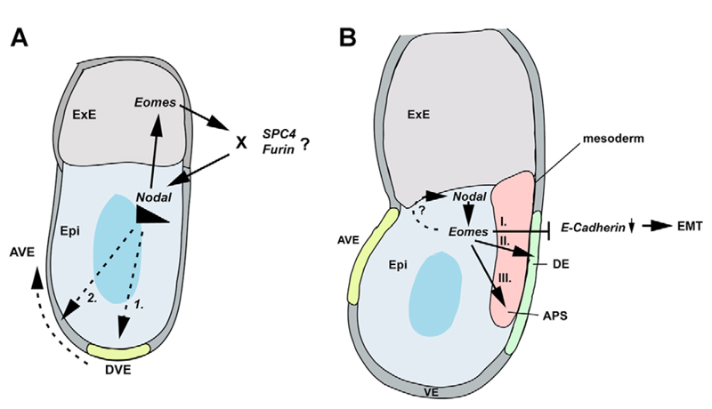
The T-box transcription factor eomesodermin (Eomes) has been implicated as an important component in germ layer induction and patterning in vertebrate embryos. In the mouse, Eomes is essential for development of the trophectoderm lineage and Eomes loss-of-function mutants arrest at implantation. Here, we have used a novel Eomes conditional allele to test Eomes functions in the embryo proper. Eomes-deficient embryos express both Fgf8 and its downstream target Snail at normal levels but surprisingly fail to downregulate E-cadherin. Eomes functional loss thus efficiently and profoundly blocks EMT and concomitant mesoderm delamination. Marker analysis as well as fate-mapping and chimera studies demonstrate for the first time that Eomes is required for specification of the definitive endoderm lineage. We also describe developmental abnormalities in Eomes/Nodal double heterozygotes, and demonstrate that these phenotypes reflect Eomes and Nodal interactions in different tissue sites. Collectively, our experiments establish that Eomes is a key regulator of anteroposterior axis formation, EMT and definitive endoderm specification in the mouse.


Figures

Fig. 1
Fig. 1
Cre mediated excision of a new Eomes conditional allele results in a null mutation. (A) To conditionally inactivate Eomes, exons 2-5 encoding the Tbox domain were flanked with LoxP sites (blue arrows) and a LoxP-flanked selectable marker (PGK.hygro) was introduced in intron 5-6. ES cell clones were screened by Southern blot on Ncol-digested DNA and the presence of the 5′ LoxP site confirmed on Xbal-digested DNA as indicated. (B) Correctly targeted clones were subjected to transient expression of Cre-recombinase. (C) HindIII digested DNA was analysed by Southern blot to detect various configurations. Green arrowheads represent the primer-sites for PCR genotyping. (D) Cultured blastocysts from Eomes N/+ intercrosses were analysed after 72 hours. Whereas wild-type and Eomes N/+ heterozygous blastocysts develop trophoblast outgrowths with typical giant cells (arrows), Eomes N/N blastocysts fail to form outgrowths (I), or display severely reduced numbers of giant cells (II). (E) Multiplex PCR genotyping of tail DNA from mice at weaning age using the primers indicated in B showing viability of Eomes CA/CA and Eomes CA/N animals. H, HindIII; Hp, HpaI; Nc, Ncol; Sa, Sad; X, Xbal.
Fig. 2
Fig. 2
Epiblast-specific Eomes deletion blocks gastrulation at the primitive streak stage. (A-F) Whole-mount in situ hybridisation with molecular markers for the AVE (Hex and Cer1) and posterior epiblast (Wnt3) at E6.5 reveal correct AP axis specification in Eomes N/CA; Sox2.Cre mutant embryos. (G-H’) At E7.5, Eomes epiblast mutants show a distinct posterior thickening of the epiblast and lack the mesodermal tissue layer as seen in histological sections. (I,J) Posterior (brachyury/Bra) and (K-N) intermediate (Mixl1, Tbx6) streak markers are expressed in Eomes epiblast mutants, but the node fails to form, as revealed by the lack of Shh expression (O,P). (Q,R) Consistent with this, scanning electron microscopy shows lack of a morphological node at the distal tip of mutant embryos (boxed areas indicate the node forming region). (S,T) Instead, the Otx2 expression domain is expanded in Eomes mutants. (U-X) At E7.5 expression of the DE marker Cer1 and the AME marker Foxa2 are lost and (Y,Z) expression of the VE marker AFP fails to become localized to the extra-embryonic region.
Fig. 3
Fig. 3
Eomes mutant epiblast cells fail to contribute to the endoderm layer. Fate-mapping analysis of the epiblast via Sox2.Cre and the ROSA26R reporter allele show that the outer endodermal layer (arrows) overlying the embryo of (A-A”) E7.75 control embryos is entirely derived from ROSA26-positive epiblast cells. (B-B”) Eomes N/CA; Sox2.Cre; ROSA26R/+ mutant epiblast cells fail to contribute to the endoderm layer (arrows). Occasionally, single lacZ-positive cells are found in the endodermal layer, most probably owing to perdurance of Cre expressed in the early ICM from the Sox2.Cre transgene.
Fig. 4
Fig. 4
Eomes-deficient epiblast exhibits normal Fgf8/Snail expression but fails to downregulate E-cadherin. (A) Scanning electron microscopy of transverse sections through E7.5 control and Eomes mutant embryos shows the mesenchymal morphology of cells accumulating at the primitive streak of mutant embryos. Eomes mutants are devoid of a mesodermal cell layer. (B) Immunofluorescence staining using an anti-E-cadherin antibody in E7.5 Eomes mutant embryos reveals failure of E-cadherin downregulation at the PS stage. Whereas mesoderm of wild-type embryos is devoid of E-cadherin (arrowhead), the distinctive tissue mass in mutants retains E-cadherin. (C,D) Fgf8 and Snail are expressed at appropriate sites and with normal intensity in Eomes N/CA; Sox2.Cre embryos when analysed by whole-mount in situ hybridisation (C) or in situ hybridisation on sections (D). The mutant PS shows overlapping expression of E-cadherin and Snail, whereas in wild-type controls the expression domains are mutually exclusive. (C) The Fgf target Spry2 is widely expressed in Eomes mutants. (E) Wild-type and Eomes mutant primitive streak explant cultures show indistinguishable migration behaviour and efficiently downregulate E-cadherin in migrating cells. Broken lines indicate the border of E-cadherin-positive explants.
Fig. 5
Fig. 5
Cell-autonomous Eomes requirements for cell migration from the primitive streak and endoderm specification. (A) Eomes-deficient ES cells were generated by retargeting the remaining wild-type allele in Eomes N/+ heterozygous ES cells. (B) Eomes N/N ES cells were introduced into ROSA26LacZ blastocysts, as depicted and chimeric embryos analysed histologically at E7.5 (C-G) and E9.5 (H-N). (C-G) /acZ-negative Eomes N/N ES cells (counterstained with Eosin) are found randomly distributed in the epiblast at E7.5 (C′,D-G) but fail to exit the PS and accumulate in cell masses in the amniotic cavity. (H) Highly chimeric embryos at E9.5 are developmentally delayed and show a relative paucity of mesoderm derivatives, resulting in gross abnormalities affecting the heart, somites, axis elongation and embryonic turning. (I-N) Eomes N/N mutant ES cells fail to contribute to definitive endoderm derivatives and cannot be found within the gut tube at any level (arrows in H-N). (N) Chimeric embryos occasionally exhibit posterior neural tube duplications (arrowheads).
Fig. 6
Fig. 6
Severely affected Eomes N/+; NodalLacZ/+ double heterozygotes fail to rotate the PD axis. (A,A’) Nodal expression normally confined to the posterior epiblast at E6.5, (B,B’) persists throughout the entire proximal epiblast of Eomes N/+ ; Nodal LacZ/+ double heterozygous embryos. (C-F) AVE-markers (Hex and Cer) remain localized to the distal tip of double heterozygous mutant embryos. (G-L) Consequently posterior marker genes (cripto, Eomes and brachyury) are expressed throughout the proximal epiblast. (M,N) Spc4 is expressed at normal levels in the ExE of mutant embryos, which fails to be displaced proximally. (O-P’) From late gastrulation stages onwards, double heterozygous mutants show gross disturbances of germ layer formation, and massive cell accumulations of mesenchymal cells within in the amniotic cavity (arrowhead in P′, lacZ staining) and severe constrictions between the embryonic and extra-embryonic regions of the embryo (arrows in P). (Q-T) At E7.5, the mesoderm marker Fgf8 is widely expressed in mutants, whereas Cer1, which marks newly formed DE, fails to be expressed.
Fig. 7
Fig. 7
A second category of Eomes N/+; Nodal LacZ/+ double heterozygous mutants fails to specify the anterior primitive streak and exhibit partial axis duplications. A proportion of compound heterozygous embryos correctly specify the AP axis, but show reduced expression of DE markers (A,B) Hex and (C-D’) Cer1 at E6.5 and E7.5, respectively. Anterior axial midline tissues are lost, as seen by (E,F) truncated brachyury expression anterior to the PS (asterisk) and (G-H’) absence of Shh (arrows indicate remaining expression in the node). (H,H’) Head folds are present at E8.0 in mutant embryos (asterisks), but (I,J) anterior neural identity and Otx2 expression is lost by E8.5. (K-N) At E9.5 double heterozygous mutants exhibit severe anterior truncations, and various degrees of heart defects, ranging from (L) looping defects (arrowheads) to (N) severe malformations with cardia bifida, while posterior development remains grossly unaffected. (O,P) A proportion of Eomes N/+ ; Nodal LacZ/+ compound heterozygous mutants show duplications of the node and notochord, as revealed by Shh expression marking both node and notochord, or (Q-R′) staining for the Nodal LacZ expression in the node. Embryos with node duplications lack Nodal expression in the left lateral plate mesoderm (arrows).
Fig. 8
Fig. 8
Model of Eomes action in gastrulation. (A) At pregastrula stages, Eomes and Nodal expression are restricted to the ExE and epiblast (Epi), respectively. Nodal signalling is required first to induce formation of the DVE, which requires elevated Nodal signalling-levels for migration to the anterior side. A positive-feedback loop between Eomes in the ExE and Nodal in the epiblast is necessary to maintain high Nodal signalling thresholds required for DVE migration. Although Nodal signalling may directly regulate Eomes in the ExE, the requirement for Eomes in maintaining Nodal levels may be indirect possibly via regulation of levels of pro-protein convertases Furin and/or SPC4 in the ExE. (B) During gastrulation, Eomes expression is initiated in the posterior proximal epiblast and the PS, where it overlaps with Nodal. Eomes participates in the transcriptional control of E-cadherin downregulation, essential for cells to traverse the PS and delaminate as mesoderm and endoderm. Additionally, Eomes is required to specify the DE lineage and acts cooperatively with Nodal signalling to activate target genes responsible for APS specification.

Similar articles

Cited by

References

    1. Batlle E, Sancho E, Franci C, Dominguez D, Monfar M, Baulida J, Garcia De, Herreros A. The transcription factor snail is a repressor of E- cadherin gene expression in epithelial tumour cells. Nat Cell Biol. 2000;2:84–89. - PubMed
    1. Beck S, Le Good JA, Guzman M, Ben Haim N, Roy K, Beermann F, Constam DB. Extraembryonic proteases regulate Nodal signalling during gastrulation. Nat Cell Biol. 2002;4:981–985. - PubMed
    1. Beddington RS, Robertson EJ. Axis development and early asymmetry in mammals. Cell. 1999;96:195–209. - PubMed
    1. Ben-Haim N, Lu C, Guzman-Ayala M, Pescatore L, Mesnard D, Bischofberger M, Naef F, Robertson EJ, Constam DB. The nodal precursor acting via activin receptors induces mesoderm by maintaining a source of its convertases and BMP4. Dev Cell. 2006;11:313–323. - PubMed
    1. Bjornson CR, Griffin KJ, Farr GH, Terashima A, Himeda C, Kikuchi Y, Kimelman D. Eomesodermin is a localized maternal determinant required for endoderm induction in zebrafish. Dev Cell. (3) 2005;9:523–533. - PubMed

Publication types

MeSH terms