. 2008 Jan 2;28(1):3-9.
doi: 10.1523/JNEUROSCI.4405-07.2008.

Alzheimer's disease (AD)-like pathology in aged monkeys after infantile exposure to environmental metal lead (Pb): evidence for a developmental origin and environmental link for AD

Affiliations

Alzheimer's disease (AD)-like pathology in aged monkeys after infantile exposure to environmental metal lead (Pb): evidence for a developmental origin and environmental link for AD

Jinfang Wu et al. J Neurosci. .

Abstract

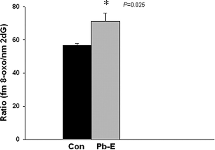
The sporadic nature of Alzheimer's disease (AD) argues for an environmental link that may drive AD pathogenesis; however, the triggering factors and the period of their action are unknown. Recent studies in rodents have shown that exposure to lead (Pb) during brain development predetermined the expression and regulation of the amyloid precursor protein (APP) and its amyloidogenic beta-amyloid (Abeta) product in old age. Here, we report that the expression of AD-related genes [APP, BACE1 (beta-site APP cleaving enzyme 1)] as well as their transcriptional regulator (Sp1) were elevated in aged (23-year-old) monkeys exposed to Pb as infants. Furthermore, developmental exposure to Pb altered the levels, characteristics, and intracellular distribution of Abeta staining and amyloid plaques in the frontal association cortex. These latent effects were accompanied by a decrease in DNA methyltransferase activity and higher levels of oxidative damage to DNA, indicating that epigenetic imprinting in early life influenced the expression of AD-related genes and promoted DNA damage and pathogenesis. These data suggest that AD pathogenesis is influenced by early life exposures and argue for both an environmental trigger and a developmental origin of AD.


Figures

Figure 1.
Figure 1.
Changes in mRNA expression of APP, BACE1, and Sp1 in the frontal association cortex of aged monkeys after developmental exposure to Pb. The frontal association cortical tissue of 23-year-old control (Con) and Pb-exposed (Pb-E) monkeys were analyzed for mRNA expression of APP, BACE1, and Sp1. The mRNA expression of APP, BACE1, and Sp1 were validated by real-time PCR. Data shown represent the mean ± SEM (4 animals in each group). Values marked with an asterisk are significantly different from their corresponding controls (p < 0.05) as determined by Student's t test.
Figure 2.
Figure 2.
Elevation of Aβ levels in the frontal association cortex of 23-year-old cynomolgus monkeys after developmental exposure to Pb. Brain tissue of control (Con) and Pb-exposed (Pb-E) 23-year-old cynomolgus monkeys was used for the analysis of Aβ levels (A, B). The levels of Aβ were measured in the frontal association cortical tissue using ELISA as described in Materials and Methods. Data shown represent the mean ± SEM for four animals in each group. Values marked with an asterisk are significantly different from their corresponding controls (p < 0.05) as determined by Student's t test.
Figure 3.
Figure 3.
Photomicrographs showing AD-like pathology in the frontal association cortex of 23-year-old cynomolgus monkeys after developmental exposure to Pb. Brain tissue of control (Con) and Pb-exposed (Pb-E) 23-year-old cynomolgus monkeys was used for the analysis of immunohistochemical analysis of AD-like pathology. Sections were prepared and stained with the Aβ-specific antibody, which recognizes both Aβ1–40 and Aβ1–42 as discussed in Materials and Methods. The arrows point to Aβ-containing plaques as well as granular and intracellular staining. The staining characteristics are similar to those reported in the literature.
Figure 4.
Figure 4.
DNA methyltransferase activity in cortical neuronal cells of mice and in monkey brains. A, B, Control and Pb-exposed mouse cortical neuronal cells (A) and frontal association cortical tissue of 23-year-old control (Con) and Pb-exposed (Pb-E) monkeys (B) were used to estimate the DNA methyltransferase activity as described in Materials and Methods. Data shown represent the mean ± SEM for four independent determinations (4 animals in each group for monkey data). Values marked with an asterisk are significantly different from their corresponding controls (p < 0.05) as determined by Student's t test.
Figure 5.
Figure 5.
Oxidative DNA damage in control and infantile-exposed aged monkey brains. Frontal association cortical tissues were obtained from 23-year-old control (Con) and Pb-exposed (Pb-E) monkeys and used to measure the marker of oxidative DNA damage as described in Materials and Methods. Data shown represent the mean ± SEM for four animals in each group. Values marked with an asterisk are significantly different from their corresponding controls (p < 0.05) as determined by Student's t test.
Figure 6.
Figure 6.
Model of effects of oxidative damage on SP1-mediated expression of APP via interference with MECP2 binding of methylated CpG dinucleotides. The APP promoter and 5′-UTR from −1000 bp to the “ATG” start codon at +148 are shown. CpG dinucleotides are indicated on the sequence, as is the +1 transcription start site. A, Undamaged DNA, showing the binding of one or more MECP2 to methylated CpG, competing against SP1. APP expression is at normal levels. B, Conversion of G residues to 8-oxo-dG or mC to 5-hydroxymethylcytosine blocks binding of some MECP2 to methylated CpG, potentially permitting additional binding of SP1 but with insufficient levels to significantly alter target gene expression levels. C, Normal changes of development, maturity, and aging result in triggering of latent SP1 regulatory alterations, which significantly increase levels of the transcription factor, permitting SP1 to outcompete remaining MECP2 binding, increasing expression of target genes.

Similar articles

Cited by

References

    1. Abdolmaleky HM, Smith CL, Faraone SV, Shafa R, Stone W, Glatt SJ, Tsuang MT. Methylomics in psychiatry: modulation of gene-environment interactions may be through DNA methylation. Am J Med Genet B Neuropsychiatr Genet. 2004;127:51–59. - PubMed
    1. Abdolmaleky HM, Cheng KH, Russo A, Smith CL, Faraone SV, Wilcox M, Shafa R, Glatt SJ, Nguyen G, Ponte JF, Thiagalingam S, Tsuang MT. Hypermethylation of the reelin (RELN) promoter in the brain of schizophrenic patients: a preliminary report. Am J Med Genet B Neuropsychiatr Genet. 2005;134:60–66. - PubMed
    1. Ball MJ, Lo P. Granulovacuolar degeneration in the ageing brain and in dementia. J Neuropathol Exp Neurol. 1977;36:474–487. - PubMed
    1. Barker DJ, Winter PD, Osmond C, Margetts B, Simmonds SJ. Weight in infancy and death from ischaemic heart disease. Lancet. 1989;2:577–580. - PubMed
    1. Basha MR, Wei W, Bakheet SA, Benitez N, Siddiqi HK, Ge YW, Lahiri DK, Zawia NH. The fetal basis of amyloidogenesis: exposure to lead and latent overexpression of amyloid precursor protein and β-amyloid in the aging brain. J Neurosci. 2005;25:823–829. - PMC - PubMed

Publication types

MeSH terms