. 2008 Jan 2;28(1):10-20.
doi: 10.1523/JNEUROSCI.3403-07.2008.

Cerebellar dysfunction explains the extinction-like abolition of conditioned eyeblinks after NBQX injections in the inferior olive

Affiliations

Cerebellar dysfunction explains the extinction-like abolition of conditioned eyeblinks after NBQX injections in the inferior olive

Svitlana Zbarska et al. J Neurosci. .

Abstract

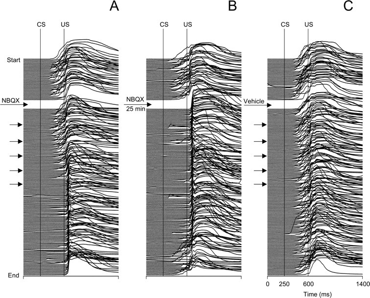
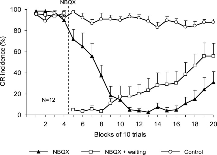
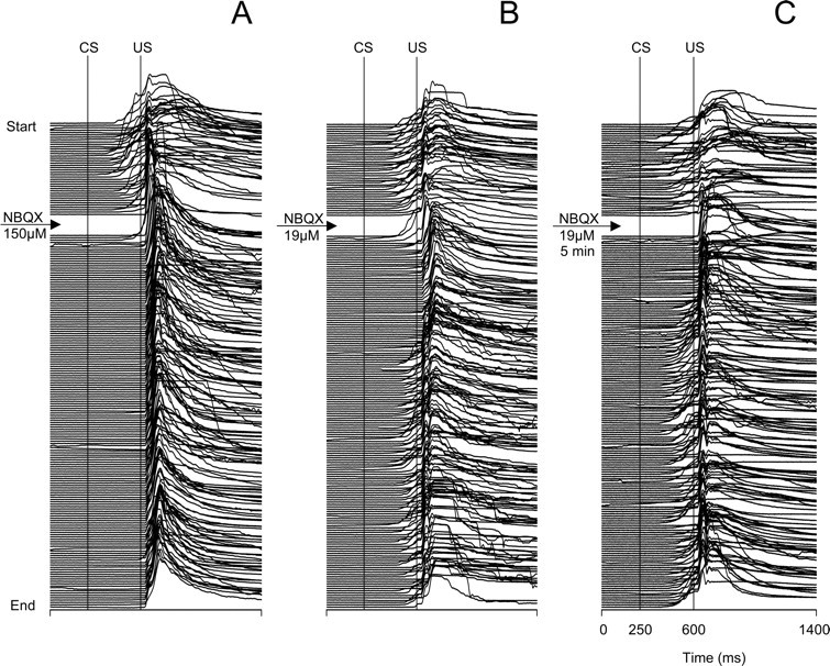
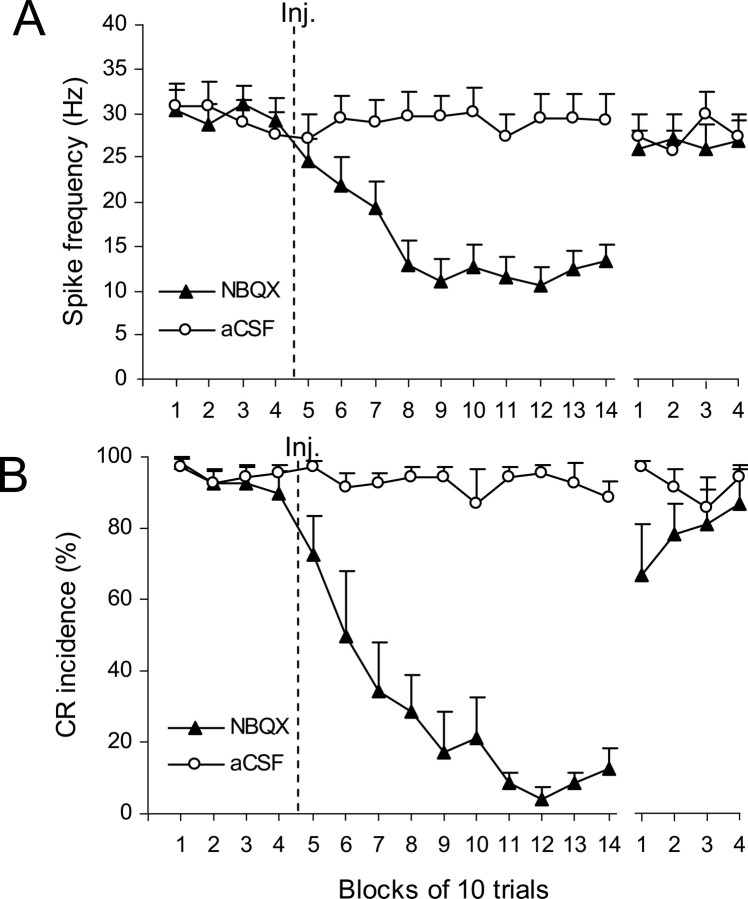
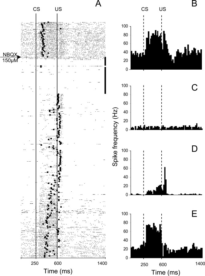
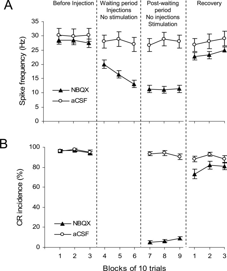
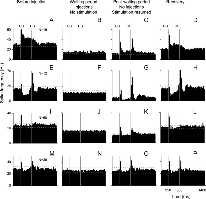
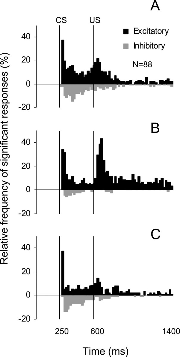
Classical conditioning of the eyeblink response is a form of motor learning that is controlled by the intermediate cerebellum and related brainstem structures. The inferior olive (IO) is commonly thought to provide the cerebellum with a "teaching" unconditioned stimulus (US) signal required for cerebellar learning. Testing this concept has been difficult because the IO, in addition to its putative learning function, also controls tonic activity in the cerebellum. Previously, it was reported that inactivation of AMPA/kainate receptors in the IO produces extinction of conditioned responses (CRs), suggesting that it blocks the transmission of US signals without perturbing the functional state of the cerebellum. However, the electrophysiological support for this critical finding was lacking, mostly because of methodological difficulties in maintaining stable recordings from the same set of single units throughout long drug injection sessions in awake rabbits. To address this critical issue, we used our microwire-based multiple single-unit recording method. The IO in trained rabbits was injected with the AMPA/kainate receptor blocker, 1,2,3,4-tetrahydro-6-nitro-2,3-dioxo-benzo[f]quinoxaline-7-sulfonamide (NBQX), and its effects on CR expression and neuronal activity in the cerebellar interposed nuclei (IN) were examined. We found that NBQX abolished CR expression and that delayed drug effects were independent of the presentation of the conditioned stimulus and were therefore not related to extinction. In parallel to these behavioral effects, the spontaneous neuronal activity and CR-related neuronal responses in the IN were suppressed, suggesting cerebellar dysfunction. These findings indicate that testing the role of IO in learning requires methods that do not alter the functional state of the cerebellum.


Figures

Figure 1.
Figure 1.
Location of injection sites in the IO injection group (•; n = 8) and in the IN recording group (★; n = 4). A–H, The identified sites in individual animals were transferred to a set of standardized coronal sections of the rabbit medulla, arranged in rostral-to-caudal order. The anterior–posterior distance in millimeters from the rostral pole of the IO is on the lower right side of each section. All injection sites were found within or close to the rostral part of the inferior olivary complex. Injections of NBQX at all marked sites abolished CR expression. Injection site labeled “1” yielded an immediate CR abolition after a 1.0 μl injection of either 150 or 19 μm NBQX. The site labeled 12 was the least effective because CR abolition required 3.5 μl of 150 μm NBQX injected over 35 min. DAO, Dorsal accessory inferior olive; IOM, medial inferior olive; IOPr, principal inferior olive; IOA, subnucleus A of the medial inferior olive; IOB, subnucleus B of the medial inferior olive; IOK, cap of Kooy of the medial inferior olive; Sp5N, spinal trigeminal nucleus; sp5, spinal trigeminal tract; Pr, prepositus hypoglossal nucleus; 7N, facial nucleus; icp, inferior cerebellar peduncle; MVe, medial vestibular nucleus; SolN, solitary nucleus; Amb, ambiguous nucleus; 12n, hypoglossal nerve; 10N, dorsal motor nucleus of the vagus; DCN, dorsal cochlear nucleus.
Figure 2.
Figure 2.
Individual examples from the same subject showing behavioral effects of microinjecting NBQX and vehicle (aCSF) into the IO. Each stack plot represents a complete printout of eyeblinks from a 200-trial injection experiment. The experiments start at the top, and each eyeblink trace represents one trial. The timing of injections is denoted by arrows in each stack plot. Conditioned eyeblinks are upward deflections of the signal between the vertical CS and US onset markers. A, Eyeblinks in an experiment where CR testing was resumed immediately after the injection of NBQX. Note that CRs were gradually abolished 23 min after NBQX. B, Stack plot of eyeblink mechanograms from an experiment in which the rabbit was injected with the same amount of drug, but the CR testing was delayed by inserting a waiting period for NBQX diffusion. In this test, CRs were absent immediately after stimuli resumed, indicating that the delayed drug effect in part A was simply a function of time, most likely related to drug diffusion. Note the partial recovery of CRs as the waiting experiment continued an extra 25 min. C, Eyeblinks in the control experiment in which injections of the same amount of vehicle (aCSF) did not affect CRs.
Figure 3.
Figure 3.
Effects of NBQX on CR incidence (±SE) in IO injection rabbits (n = 12) when tested immediately after the injection (▴) and after the diffusion waiting period (□) are compared with those observed after the control injection of vehicle (○). The vertical dashed line denotes the onset of NBQX injections. Note that up to six blocks of trials were required to obtain complete CR abolition in the nonwaiting test, whereas CRs were immediately absent in the first postinjection block of the waiting test. Injection of vehicle (aCSF) did not affect CR incidence.
Figure 4.
Figure 4.
Stack plots of all eyeblinks recorded in three separate experiments from the same animal showing the behavioral effects of microinjecting different concentrations of NBQX in the IO. A, An experiment where CRs were tested immediately after an injection of 1.0 μl of NBQX (150 μm). CRs were almost immediately abolished after 1 min of postinjection training. B, Eyeblinks in an experiment where 1.0 μl of eight times less concentrated NBQX (19 μm) was injected and then an immediate test was performed. CRs were abolished after several postinjection trials, but the duration of the effect was shorter than that obtained with the higher concentration of NBQX shown in A. C, Eyeblinks in an experiment where postinjection testing of 19 μm NBQX was delayed by a 5 min diffusion waiting period. Note that CRs were absent starting from the first postinjection trial, and then 10 min later they started to recover. For additional plot information, see Figure 2.
Figure 5.
Figure 5.
Reconstruction of cell recording sites in the IN. Of 12 electrode bundles in four rabbits, eight were located in the anterior IN and four at the caudal border of the anterior interposed nucleus (InA) between the dentate nucleus (DN) and posterior interposed nucleus (InP). Their position was transferred to a set of standardized coronal sections of the rabbit cerebellar nuclear region. A–D, Sections are arranged in rostral-to-caudal order. LV, lateral vestibular nucleus; SV, superior vestibular nucleus; FN, fastigial nucleus; scp, superior cerebellar peduncle; icp, inferior cerebellar peduncle.
Figure 6.
Figure 6.
Group effects of NBQX on CR performance and on IN activity during the nonwaiting test. The last four blocks of trials represent the recovery period and are separated from the rest of the plot by a gap. A, Effects of NBQX on mean baseline activity (±SE) of IN cells (n = 27). The firing rate of IN cells gradually decreased within the first four blocks of trials after the injection of NBQX (▴), and it remained at slightly above 10Hz for the next 60 trials. The spontaneous activity almost completely recovered during the last four blocks of the experiment. Injections of aCSF (○) had no effect on IN firing rate (n = 20). B, Effects of NBQX (▴) on mean CR incidence (±SE; n = 7) in the same set of experiments as depicted in A. CRs were gradually abolished and then partially recovered by the end of testing. This behavioral effect parallels changes in IN activity shown in A. Control injections of vehicle did not affect CR performance.
Figure 7.
Figure 7.
An example from a waiting period test of the parallel effects of NBQX on CR performance and on the activity of a task-modulated IN cell. This experiment consisted of 260 trials with the two 0.5 μl NBQX injections administered during the 40-trial no-stimulation period that began immediately after 40 preinjection trials. A, Raster plot of IN cell activity during the same experiment. The experiment starts at the top with each row representing one trial, and each dot marking the occurrence of an action potential. The black square on each row corresponds to the onset of the eyeblink in that particular trial. The 40 black squares at the ends of rows correspond to the no-stimulation waiting period. This cell responded with excitation during the CS–US interval followed by a combined excitatory/inhibitory response to the US. Shortly after the first NBQX injection, the firing rate of this cell's activity precipitously declined. When stimulation was resumed, CRs were abolished immediately, baseline activity remained suppressed, and modulation during the CS–US interval was severely reduced whereas the relative excitatory modulation to the US became more distinct. The neuronal activity gradually recovered toward the end of the experiment in parallel with the recovery of behavioral CRs. B–E, Peristimulus histograms of the same IN unit constructed for 40 trials before the injection (B), for 40 postinjection waiting trials when stimulation was paused (C), for 40 postwaiting period trials when stimulation was resumed (D), and for the last 40 trials of the experiment (E). Bin width for histograms in B–E is 20 ms. CS, Onset of conditioned stimulus; US, onset of unconditioned stimulus.
Figure 8.
Figure 8.
Group effects of NBQX on CR performance and on IN activity during the waiting test. Each period of the experiment is represented by three consecutive blocks of 10 trials: before injection, no-stimulation waiting period, postwaiting period with stimulation, and recovery. Periods are separated by vertical dashed lines. If the waiting period was longer than 30 trials, then the last 30 trials before stimulation was resumed were included in the plot. A, Effects of NBQX (▴) on mean baseline activity (±SE; n = 88) of IN cells. The baseline firing rate of IN cells gradually decreased during the waiting period, it remained low when stimulation was resumed and it partially recovered during the last three blocks of the experiment. Injecting aCSF (○) had no significant effect on IN firing rate (n = 36). B, Effects of NBQX (▴) on mean CR incidence (±SE; n = 32) in the same set of experiments as depicted in A. CRs were completely abolished when stimulation was resumed and then partially recovered by the end of testing. This behavioral effect parallels changes in IN activity shown in A. Control injections of vehicle (○) did not affect CR performance.
Figure 9.
Figure 9.
Effects of NBQX and aCSF injections on cell activity modulation. Mean peristimulus histograms are arranged in rows corresponding to cell types and in columns representing the activity before injection, during the no stimulation waiting period, postwaiting period when stimulation was resumed, and recovery. AD, Cells exhibiting predominantly excitatory responses (n = 16). E–H, Cells that were inhibited during the CS–US interval (n = 12). I–L, Nonmodulated cells (n = 60). M–P, Cells recorded during control injections of vehicle (n = 36). CS, Conditioned stimulus onset; US, Unconditioned stimulus onset. Bin width is 20 ms. Horizontal lines in each histogram represent tolerance limits. For a more detailed description, see Results, Effect of NBQX injection in the IO on neuronal activity in the IN.
Figure 10.
Figure 10.
A–C, Frequency histograms of the incidence of significant excitatory (black bars) and inhibitory (gray bars) responses in the population of 88 cells recorded during the NBQX waiting experiments (A, before injection; B, after injection when stimulation was resumed; C, during last 30 trials of experiments). The fraction of cells exhibiting long-latency excitatory or inhibitory responses in the CS–US interval decreased after the NBQX application. Also, the incidence of short-latency excitation to the CS remained unchanged and the incidence of excitatory responses to the US increased after NBQX. These effects tended to recover toward the end of experiments.

Similar articles

Cited by

References

    1. Aksenov D, Serdyukova N, Irwin K, Bracha V. GABA neurotransmission in the cerebellar interposed nuclei: involvement in classically conditioned eyeblinks and neuronal activity. J Neurophysiol. 2004;91:719–727. - PubMed
    1. Batini C, Billard JM, Daniel H. Long-term modification of cerebellar inhibition after inferior olive degeneration. Exp Brain Res. 1985;59:404–409. - PubMed
    1. Benedetti F, Montarolo PG, Strata P, Tempia F. Inferior olive inactivation decreases the excitability of the intracerebellar and lateral nuclei in the rat. J Physiol (Lond) 1983;340:195–208. - PMC - PubMed
    1. Bengtsson F, Svensson P, Hesslow G. Feedback control of Purkinje cell activity by the cerebello-olivary pathway. Eur J Neurosci. 2004;20:2999–3005. - PubMed
    1. Bracha V, Webster ML, Winters NK, Irwin KB, Bloedel JR. Effects of muscimol inactivation of the cerebellar interposed-dentate nuclear complex on the performance of the nictitating membrane response. Exp Brain Res. 1994;100:453–468. - PubMed

Publication types

MeSH terms

Substances

LinkOut - more resources