. 2008 Jan 2;28(1):177-88.
doi: 10.1523/JNEUROSCI.3573-07.2008.

L1 interaction with ankyrin regulates mediolateral topography in the retinocollicular projection

Affiliations

L1 interaction with ankyrin regulates mediolateral topography in the retinocollicular projection

Mona Buhusi et al. J Neurosci. .

Abstract

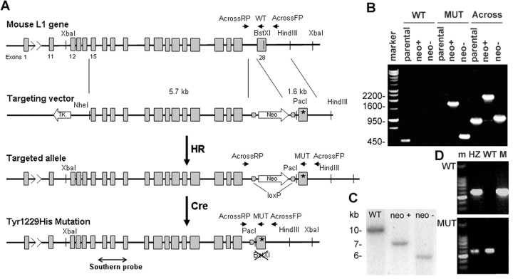
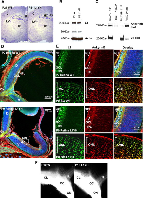
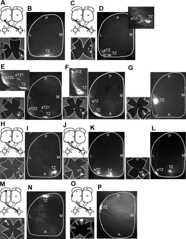
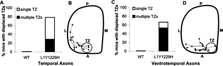
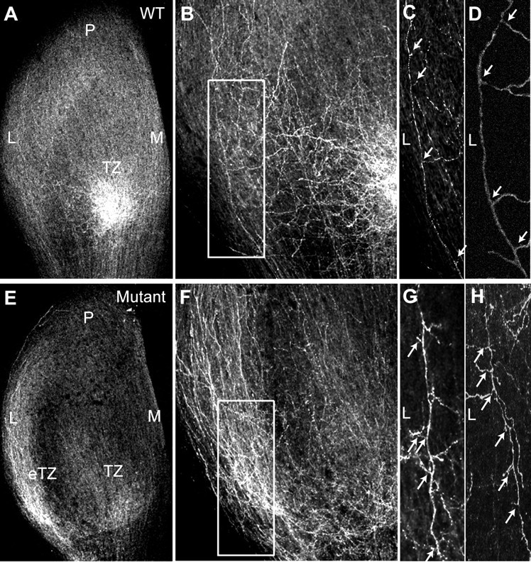
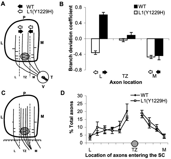
Dynamic modulation of adhesion provided by anchorage of axonal receptors with the cytoskeleton contributes to attractant or repellent responses that guide axons to topographic targets in the brain. The neural cell adhesion molecule L1 engages the spectrin-actin cytoskeleton through reversible linkage of its cytoplasmic domain to ankyrin. To investigate a role for L1 association with the cytoskeleton in topographic guidance of retinal axons to the superior colliculus, a novel mouse strain was generated by genetic knock-in that expresses an L1 point mutation (Tyr1229His) abolishing ankyrin binding. Axon tracing revealed a striking mistargeting of mutant ganglion cell axons from the ventral retina, which express high levels of ephrinB receptors, to abnormally lateral sites in the contralateral superior colliculus, where they formed multiple ectopic arborizations. These axons were compromised in extending interstitial branches in the medial direction, a normal response to the high medial to low lateral SC gradient of ephrinB1. Furthermore, ventral but not dorsal L1(Y1229H) retinal cells were impaired for ephrinB1-stimulated adhesion through beta1 integrins in culture. The retinocollicular phenotype of the L1(Tyr1229His) mutant provides the first evidence that L1 regulates topographic mapping of retinal axons through adhesion mediated by linkage to the actin cytoskeleton and functional interaction with the ephrinB/EphB targeting system.


Figures

Figure 1.
Figure 1.
Generation of the L1(Y1229H) knock-in mouse. A, Schematic representation of the genetic modification at the L1 locus: wild-type exon 28 was replaced by a modified exon 28 in which the codon corresponding to 1229Tyr was changed to encode His by homologous recombination (HR) in ES cells; the neomycin-resistance gene introduced at the same time in intron 27, was subsequently removed by cre-mediated recombination in ES cells (Cre). B, PCR analysis of ES clones (parental ES cells, neomycin-positive clone after HR, neomycin-negative clone after cre recombination; marker, 1 kb DNA ladder). Lanes 1–3, PCR for the wild-type allele, using the WT and AcrossL1RP primers. Lanes 4–6, PCR for the mutant allele, using the MUT and AcrossL1RP primers. Lanes 7–9, PCR covering the region modified by recombination, using the AcrossL1 FP and RP primers. C, Southern blot screening of recombined neomycin-positive clones and neomycin-negative clones after cutting with XbaI and PacI (site introduced through recombination), using a probe corresponding to exons 16–19. D, Mouse genotyping by PCR using allele specific primers: top row, MUT and AcrossL1RP; bottom row, WT and AcrossL1RP; m, marker (100 bp DNA ladder); HZ, heterozygote female; WT, wild-type male; M, hemizygous L1YH.
Figure 2.
Figure 2.
L1 expression and colocalization with ankyrinB in brain and retina of WT and L1(Y1229H) mice. A, Nissl staining of WT and L1(Y1229H) mice at P21 (strain 129). C, Cortex; HC, hippocampus; TH, thalamus; LV, lateral ventricle; cc, corpus callosum. B, Western blot analysis of P3 brain lysates (45 μg) from WT (lane 1) and mutant (lane 2) mice using an L1 antibody. C, Coimmunoprecipitation of ankyrinB with L1 from WT mouse superior colliculus lysates (P8). No ankyrinB coimmunoprecipitated with nonimmune mouse IgG or with L1(Y12229H) protein. D, Immunofluorescence localization of L1 and ankyrinB in sagittal sections of the P0 mouse retina. E, Localization of L1 and ankyrinB in sagittal sections of the P0 mouse retina (high magnification) and in coronal sections of the superior colliculus in the anterior third of the anteroposterior axis: L1, green; ankyrinB, red; Nissl stain, pseudocolored blue. NFL, Nerve fiber layer; GCL, ganglion cell layer; IPL, inner plexiform layer; D, dorsal; V, ventral; ON, optic nerve; ONL, optic nerve layer in the SC. F, Optic chiasm visualized by DiI tracing from the left retina. Contralateral (CL) and ipsilateral (IL) projections in WT and L1(Y1229H) littermates were of similar sizes.
Figure 3.
Figure 3.
Defects in retinocollicular mapping in L1(Y1229H) mutant mice. A, B, Injection of DiI into the peripheral temporal retina (shown in flatmount) at P8 labeled a single TZ in the anterior SC in P10 WT mice. C–G, DiI injections in the peripheral temporal retina of L1(Y1229H) mutant mice revealed ectopic TZs (eTZ), displaced to lateral and slightly posterior positions in the SC. D–F, Higher magnification of the TZs (top left) revealed branches extending laterally or posteriorly toward the eTZs. H, I, Focal injections of DiI in the ventrotemporal (VT) retina labeled a single dense TZ in the anteromedial corner of the SC in P10 WT mice. J–L, Similar injections in L1(Y1229H) mutant mice resulted in eTZs displaced laterally within the anterior SC. M, N, Nasal retinal injections in L1(Y1229H) mutant mice resulted in labeling of a single TZ in the posterior SC at P10. O, P, Injection of DiI in the peripheral dorsal retina at P8 labeled a single TZ in the lateral SC in P10 L1(Y1229H) mice. L, Lateral; M, medial; A, anterior; P, posterior; D, dorsal; V, ventral; N, nasal; T, temporal.
Figure 4.
Figure 4.
Displacement of termination zones of RGC axons in L1(Y1229H) mutant mice. A, Percentage of mice with displaced TZs in the SC after temporal retinal injections at P10–P12. B, Schematic representation of the location of TZs after temporal retinal injections in L1(Y1229H) mutant mice at P10. The centers of TZs and eTZs from mutant mice (n = 10) are marked and connected. Range of location of TZs of WT mice in the anteromedial SC is depicted by the gray circle. C, Percentage of mutant mice with displaced TZs after ventrotemporal retinal injections at P10. D, Schematic representation of the aberrant eTZs after ventrotemporal retinal injections in mutant mice (n = 6). L, Lateral; M, medial; A, anterior; P, posterior.
Figure 5.
Figure 5.
Abnormal directional branch extension in WT and L1(Y1229H) mutant mice at P2. A, B, Labeling of RGC axons at P2 after focal injections of DiI in the ventrotemporal retina of WT mice show interstitial branches extending toward the nascent TZ in the anteromedial SC. C, D, Most of the branches extending from axons that navigate lateral to the future TZ are oriented medially as shown in a higher magnification of the boxed area in B from confocal z-stacks (arrows). E, F, DiI labeling of VT axons in the SC of L1(Y1229H) mice at P2 reveal an accumulation at an inappropriate ectopic TZ (eTZ) as well as at a more appropriate future TZ. G, H, Many interstitial branches of mutant VT axons are abnormally oriented toward the lateral SC, as shown in a higher magnification of the boxed area in F from z-stacks (arrows). M, Medial; L, lateral; P, posterior.
Figure 6.
Figure 6.
Branching and axonal positioning patterns in the SC of WT and L1(Y1229H) mice at P2. A, Schematic representation of directed branch extension along the mediolateral axis. The SC was divided in three bins [lateral (L), TZ, medial (M)] separated by dash lines, in relation to the forming TZ (gray circle). The orientation of each branch was recorded and graphed by bin. B, Distribution of branches in P2–P3 WT and L1(Y1229H) mutant mice after VT retinal injections. Arrows represent the direction of the branching preference in each region. The results were expressed as a branch directional coefficient as described (Hindges et al., 2002), calculated for each subject for each of the three bins of the SC as the difference in the number of medially oriented branches minus the number of laterally oriented branches, divided by the total number of branches. C, Schematic representation of RGC axon positioning in the SC in WT and mutant mice. The SC was divided into 10 segments along the mediolateral axis at the anterior border, and all labeled RGC axons within each bin were counted and represented relative to the position of the developing TZ for each injection. D, Distribution of labeled axons along the mediolateral SC axis (expressed as percentage of total) in L1(Y1229H) mice compared to WT littermates at P2, after VT retinal injections. Error bars indicate SEM.
Figure 7.
Figure 7.
L1/ankyrin interaction promotes ephrinB1-stimulated, integrin-mediated retinal cell adhesion. A, Retinal cell adhesion to fibronectin was quantified by determining the mean number of cells per field from three or more independent assays. Adhesion of WT ventral retinal cells to fibronectin was greater than WT dorsal retinal cells, and was stimulated by ephrinB1-Fc. Adhesion of L1(Y1229H) ventral retinal cells was lower than WT ventral cells and was not stimulated by ephrinB1-Fc. Total cell numbers were as follows: WT ventral retina, n = 100; WT dorsal retina, n = 80; LIYH ventral retina, n = 100; LIYH dorsal retina, n = 60. *p < 0.05, statistical significance was analyzed using the two-tailed t test. B, Echistatin peptide (5 μg/ml), an inhibitor of RGD-binding integrins, decreased adhesion of WT ventral retinal cells to fibronectin-coated dishes treated with preclustered ephrinB1-Fc fusion protein (1 μg/ml) or Fc control protein. A representative experiment (from a total of 3 experiments) is shown with n = 20 cells per condition. Statistical significance was analyzed using the two-tailed t test, *p < 0.05.

References

    1. Ango F, di Cristo G, Higashiyama H, Bennett V, Wu P, Huang ZJ. Ankyrin-based subcellular gradient of neurofascin, an immunoglobulin family protein, directs GABAergic innervation at Purkinje axon initial segment. Cell. 2004;119:257–272. - PubMed
    1. Birgbauer E, Cowan CA, Sretavan DW, Henkemeyer M. Kinase independent function of EphB receptors in retinal axon pathfinding to the optic disc from dorsal but not ventral retina. Development. 2000;127:1231–1241. - PubMed
    1. Brakebusch C, Fassler R. beta 1 integrin function in vivo: adhesion, migration and more. Cancer Metastasis Rev. 2005;24:403–411. - PubMed
    1. Burgoon MP, Grumet M, Mauro V, Edelman GM, Cunningham BA. Structure of the chicken neuron-glia cell adhesion molecule, Ng-CAM: origin of the polypeptides and relation to the Ig superfamily. J Cell Biol. 1991;112:1017–1029. - PMC - PubMed
    1. Chan W, Kordeli E, Bennett V. 440-kD ankyrinB: structure of the major developmentally regulated domain and selective localization in unmyelinated axons. J Cell Biol. 1993;123:1463–1473. - PMC - PubMed

Publication types

MeSH terms

LinkOut - more resources