. 2008 Jan 2;28(1):315-24.
doi: 10.1523/JNEUROSCI.2699-07.2008.

p75 neurotrophin receptor mediates neuronal cell death by activating GIRK channels through phosphatidylinositol 4,5-bisphosphate

Affiliations

p75 neurotrophin receptor mediates neuronal cell death by activating GIRK channels through phosphatidylinositol 4,5-bisphosphate

Elizabeth J Coulson et al. J Neurosci. .

Abstract

The pan neurotrophin receptor p75(NTR) signals programmed cell death both during nervous system development and after neural trauma and disease in the adult. However, the molecular pathways by which death is mediated remain poorly understood. Here, we show that this cell death is initiated by activation of G-protein-coupled inwardly rectifying potassium (GIRK/Kir3) channels and a consequent potassium efflux. Death signals stimulated by neurotrophin-mediated cleavage of p75(NTR) activate GIRK channels through the generation and binding of phosphatidylinositol 4,5-bisphosphate [PtdIns(4,5)P2/PIP2] to GIRK channels. Both GIRK channel activity and p75(NTR)-mediated neuronal death are inhibited by sequestration of PtdIns(4,5)P2 and application of GIRK channel inhibitors, whereas pertussis toxin treatment has no effect. Thus, p75(NTR) activates GIRK channels without the need for G(i/o)-proteins. Our results demonstrate a novel mode of activation of GIRK channels, representing an early step in the p75(NTR)-mediated cell death pathway and suggesting a function for these channels during nervous system development.


Figures

Figure 1.
Figure 1.
Chopper domain peptide-induced cell death is inhibited by potassium channel blockers A, p75NTR undergoes regulated intramembrane proteolysis, whereby the full-length p75NTR is cleaved by metalloprotease (MMP) activity within the extracellular domain. This cleavage can be induced by neurotrophic ligands such as NGF. After extracellular cleavage, the remaining CTF of p75NTR is cleaved intracellularly by presenilin-dependent γ-secretase activity, releasing the soluble intracellular domain fragment (ICD). A membrane-linked peptide corresponding to the intracellular juxtamembrane Chopper domain was used to constitutively induce neuronal death. Neuronal survival after 2 h of treatment with the death-inducing Chopper domain (black bars) or control (white bars) peptides in the presence of the K+ channel inhibitors TEA (B) and bupivacaine (C). The gray bar indicates untreated neurons (n = 3 replica wells; mean ± SD; representative of 3 and 2 experiments, respectively). D, Neuronal survival was determined after 2 h of treatment with Chopper domain or control peptides in the presence of tertiapin (n = 3 replica wells; mean ± SD; representative of 4 experiments). *p < 0.05, **p < 0.01, relative to vehicle-treated condition.
Figure 2.
Figure 2.
Extracellular cleavage of p75NTR and potassium efflux through GIRK channels is necessary for cell death. A, Summary data showing caspase activity (see Materials and Methods) in HEK293 cells transfected as indicated and treated overnight with 400 nm PMA to induce extracellular cleavage. Coexpression of functional GIRK channels with full-length p75NTR induces caspase activity above that of controls. Coexpression of the nonfunctional chimeric GIRK2-IRK1 subunit (G2IRK) instead of wild-type GIRK2 precludes the increase in caspase activity. Coexpression of the p75-Nglycos mutant (which cannot be cleaved extracellularly) with GIRK1/2 channels similarly fails to stimulate caspase activity. The CTF form of p75NTR induced caspase activity above that induced by the control or wild-type p75NTR only when coexpressed with functional GIRK1/2 channels (mean ± SEM; n = 3 replica wells; representative of 3 independent experiments). **p < 0.01, ***p < 0.001. B, Summary data showing death of HEK293T cells transfected with p75-CTF alone, or p75-CTF together with GIRK1 and either GIRK2 or a chimeric GIRK2-182-IRK1 channel (G2IRK) relative to that of cells transfected with EGFP alone (0%) (mean ± SEM; n ≥ 8). ***p < 0.001, relative to both p75-CTF alone and GIRK1/2 alone (data not shown). C, The amount of 86Rb released from transfected HEK293T cells over 60 min was measured to determine the relative efflux of potassium. Activation of GIRK channels by either βγ subunits (circles, top plot) or p75-CTF (triangles, middle plot) caused a similar level of efflux, which was significantly greater than that seen with EGFP-transfected cells (squares, bottom plot) (mean ± SD; n = 3). D, Effect of potassium concentration on apoptosome activation was measured in HEK293T cell lysates by the relative extent of caspase activity as described in Materials and Methods. Potassium concentrations >110 mm significantly inhibited apoptosome activity (n = 3 experiments; p < 0.05).
Figure 3.
Figure 3.
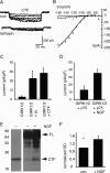
The C-terminal fragment of p75NTR activates GIRK channels. HEK293T cells were transfected with combinations of GIRK 1 and 2 subunits, βγ subunits, and CTF-p75 together with GFP for identification of positive cells (see Materials and Methods). Forty-eight hours after transfection, whole-cell recordings were made from GFP-positive HEK293T cells to ascertain GIRK channel activity. A, Inward currents evoked by hyperpolarizing voltage steps from a holding potential of −60 mV were present in HEK293T cells transfected with GIRK1/2 and p75-CTF. The p75-CTF activated inward current was blocked by tertiapin (200 nm). The steady state current–voltage relationship before and after addition of tertiapin is shown in B. C, Summary of data from currents recorded at −140 mV in cells expressing only GIRK1/2, GIRK1/2, and βγ subunits or GIRK1/2 and p75-CTF. D, Summary of data from currents recorded in HEK293 cells expressing GIRK1/2 together with full-length p75NTR with or without overnight treatment with NGF. NGF treatment stimulated a threefold increase in p75NTR-mediated GIRK channel activity. E, Representative Western blot of HEK293 cells expressing CTF (left lane), full-length p75NTR with (right lane) or without (middle lane) overnight treatment with NGF. F, Significant accumulation of the C-terminal fragment generated from full-length p75NTR was promoted by NGF treatment (mean ± SD; n = 4; **p < 0.01). con, Control.
Figure 4.
Figure 4.
p75NTR activates GIRK channels independent of G-proteins. A, Survival of neurons pretreated overnight with pertussis toxin and subsequently treated for 2 h with Chopper or control peptides. No inhibition of Chopper-mediated cell death was observed (mean ± SD; n = 3). B, Representative current–voltage relationship of HEK293T cells transfected with or without PH-PLCδ domain plasmid (PH) in combination with GIRK 1 and 2 subunits and CTF. Forty-eight hours after transfection, whole-cell recordings were made from EGFP-positive HEK293T cells in 30 mm extracellular potassium solution. The PH-PLCδ domain significantly reduced the CTF-mediated GIRK channel activity. C, Summary data for steady-state currents evoked at −90 mV for control (Con) (n = 12) cells and those transfected with PH-PLCδ (PH; n = 7).
Figure 5.
Figure 5.
p75NTR stimulates increased production of PtdIns(4,5)P2. A, Confocal images of HEK293T cells coexpressing PH-PLCδ-EYFP and either β-galactosidase (right) or p75NTR (left). B, HEK293T cells expressing p75NTR had significantly higher levels of membrane-localized EYFP. HEK293T cells expressing a noncleavable form of p75NTR (Nglycos) had significantly lower levels of membrane-localized EYFP (mean ± SD; *p < 0.01). C, Cos7 cells expressing EGFP, DN-Rac, p75NTR, or p75NTR and DN-Rac were labeled with [γ-32P]-orthophosphate, and cellular lipids were extracted and separated on oxalate-treated TLC plates before exposure to x-ray film or phosphoimage plates. D, The significant increase in generation of PtdIns(4,5)P2 (PIP2) by p75NTR was inhibited by blocking Rac activity (mean ± SD; n = 3; *p < 0.05).
Figure 6.
Figure 6.
p75NTR-mediated DRG neuronal death requires potassium flux though GIRK channels. A, Survival of DRG neurons 16 h after microinjection with GFP or p75-CTF alone or together with pGIRK2-AAA or pGIRK2/IRK expression constructs after 24 h in NGF. Expression of these subunits in the absence of death signaling had no effect on survival but prevented p75-CTF-mediated neuronal death (mean ± SD; n ≥ 3; **p < 0.01). B, Eyes of E4.5 chicks were injected with 1 μl of water containing the potassium channel inhibitor tertiapin. After 20 h, the retinal ganglion cell layer was assayed for caspase activity (see Materials and Methods). Tertiapin (50 pmol) reduced the activity significantly (**p = 0.0011). The number of retinas is indicated per condition (representative of 3 experiments).
Figure 7.
Figure 7.
Model of the proposed p75NTR- and GIRK channel-mediated cell death signaling pathway. Death signaling is activated by metalloprotease cleavage of p75NTR after stimulation by mature or proneurotrophins (NGF). The C-terminal fragment of p75NTR activates Rac, which in turn activates PIPKI, with signal transduction assisted by colocalization of signaling molecules within lipid raft-like membrane domains. The locally generated PtdIns(4,5)P2 (PIP2) then binds to the C-terminal domain of neighboring GIRK2 subunits, activating GIRK1/2 heterotrimeric channels. The resulting efflux of potassium through the GIRK channels lowers the intracellular potassium concentration, releasing inhibition of the apoptosome, and resulting in capsase activation and ultimately cell death.

References

    1. Bamji SX, Majdan M, Pozniak CD, Belliveau DJ, Aloyz R, Kohn J, Causing CG, Miller FD. The p75 neurotrophin receptor mediates neuronal apoptosis and is essential for naturally occurring sympathetic neuron death. J Cell Biol. 1998;140:911–923. - PMC - PubMed
    1. Barker PA, Barbee G, Misko TP, Shooter EM. The low affinity neurotrophin receptor, p75LNTR, is palmitoylated by thioester formation through cysteine 279. J Biol Chem. 1994;269:30645–30650. - PubMed
    1. Cain K, Langlais C, Sun XM, Brown DG, Cohen GM. Physiological concentrations of K+ inhibit cytochrome c-dependent formation of the apoptosome. J Biol Chem. 2001;276:41985–41990. - PubMed
    1. Carpenter CL, Tolias KF, Van Vugt A, Hartwig J. Lipid kinases are novel effectors of the GTPase Rac1. Adv Enzyme Regul. 1999;39:299–312. - PubMed
    1. Chen SC, Ehrhard P, Goldowitz D, Smeyne RJ. Developmental expression of the GIRK family of inward rectifying potassium channels: implications for abnormalities in the weaver mutant mouse. Brain Res. 1997;778:251–264. - PubMed

Publication types

MeSH terms