. 2008 Mar;19(3):1032-45.
doi: 10.1091/mbc.e07-05-0499. Epub 2008 Jan 2.

Ypi1, a positive regulator of nuclear protein phosphatase type 1 activity in Saccharomyces cerevisiae

Affiliations

Ypi1, a positive regulator of nuclear protein phosphatase type 1 activity in Saccharomyces cerevisiae

Jennifer P Bharucha et al. Mol Biol Cell. 2008 Mar.

Abstract

The catalytic subunit of protein phosphatase type 1 (PP1) has an essential role in mitosis, acting in opposition to the Ipl1/Aurora B protein kinase to ensure proper kinetochore-microtubule interactions. However, the regulatory subunit(s) that completes the PP1 holoenzyme that functions in this capacity is not known. We show here that the budding yeast Ypi1 protein is a nuclear protein that functions with PP1 (Glc7) in this mitotic role. Depletion of cellular Ypi1 induces mitotic arrest due to activation of the spindle checkpoint. Ypi1 depletion is accompanied by a reduction of nuclear PP1 and by loss of nuclear Sds22, a Glc7 binding partner that is found in a ternary complex with Ypi1 and Glc7. Expression of a Ypi1 variant that binds weakly to PP1 also activates the spindle checkpoint and suppresses the temperature sensitivity of an ipl1-2 mutant. These results, together with genetic interactions among YPI1, GLC7, and SDS22 mutants, indicate that Ypi1 and Sds22 are positive regulators of the nuclear Glc7 activity that is required for mitosis.


Figures

Figure 1.
Figure 1.
Depletion of Ypi1 results in G2 cell cycle delay and activation of the spindle assembly checkpoint. (A) Immunoblot analysis with anti-HA antibody of 3HA-Ypi1 in extracts from WT (KT2422) and PGAL1-3HA-YPI1 (KT2432) strains, after shifting cells from YPGal to YPD medium as described in Materials and Methods. Pgk1 serves as a loading control. (B) Growth curves of the WT (JB289-1A), PGAL1-3HA-YPI1 (JB284-2C), mad1Δ (JB289-1C), and PGAL1-3HA-YPI1 mad1Δ (JB289-4B) strains after the shift to glucose. (C) Flow cytometry analysis of DNA content for the strains in B at 12, 16, and 20 h in YPD. (D) Tub1-GFP fluorescence in WT (JB289-1A) and PGAL1-3HA-YPI1 (JB284-2C) cells after 16 h in YPD medium. Bar, 5 μm. (E) GFP-lacI fluorescence in the WT (JB338-3A), PGAL1-3HA-YPI1 (KT2668), mad1Δ (JB338-14D), and PGAL1-3HA-YPI1 mad1Δ (JB338-13B) strains at 12 h in YPD. DIC and GFP images are shown. White arrow, a large-budded cell with no spot; white arrowhead, a large-budded cell with no spot in the mother and two spots in the daughter cell.
Figure 2.
Figure 2.
Ypi1 is required for the nuclear localization of Glc7. (A) Immunoblot analysis of extracts from WT (KT1112), GLC7-mYFP (KT2422), and SDS22-mYFP (JB320-1B) strains with anti-GFP antibody. Pgk1 serves as a loading control. (B) Subcellular distribution of Glc7-mYFP in WT (KT2771) and PGAL1-3HA-YPI1 (KT2767) strains at the indicated time after shifting to YPD. Cells were examined by fluorescence microscopy with a GFP filter set for Glc7-mYFP and a rhodamine filter set for Pom34-mRFP. Bar, 5 μm. (C) Quantitative analysis of the ratio of nuclear to cytoplasmic levels of Glc7-mYFP in the WT (KT2422) (□ and ○) and PGAL1-3HA-YPI1 (KT2432; ■ and •) cells shown in B. Squares and circles indicate data from two separate experiments. To accurately compare the changes in fluorescence levels between experiments we determined the nuclear/cytoplasmic fluorescence ratio rather than the absolute signal, which can fluctuate with changes in Hg-Arc intensity. More than 50 cells were examined for each strain at each time point. Bright fluorescent punctae were excluded from the analysis. (D) Immunoblot analysis with anti-GFP antibody of Glc7-mYFP in extracts from the strains in B. Left and right panels, lane 1, GLC7-mYFP strain (KT2422); lane 2, GLC7-mYFP PGAL1-3HA-YPI1 strain (KT2432); and lane 3, WT strain (KT1112). Pgk1 serves as a loading control.
Figure 3.
Figure 3.
Ypi1 is required for the nuclear localization of Sds22. (A) Subcellular distribution of Sds22-mYFP in WT (JB328-12C) and PGAL1-3HA-YPI1 (JB331-3A) strains at the indicated times in YPD. Pom34-mRFP serves as a nuclear periphery marker. White arrows indicate loss of Sds22 from the nucleus. Cells were examined by fluorescence microscopy with a YFP filter set for Sds22-mYFP and a rhodamine filter set for Pom34-mRFP. Bar, 5 μm. (B) Immunoblot analysis of Sds22-mYFP in extracts from the strains in A. Left and right panels, lane 1, SDS22-mYFP strain (JB328-12C); lane 2, SDS22-mYFP PGAL1-3HA-YPI1 strain (JB331-3A). The Sds22 protein level at the 16-h time point is inconsistently low.
Figure 4.
Figure 4.
Variation in nuclear Glc7 levels between Glc7-RFP variants. (A) Yeast strains KT2729 (GLC7-tdimer2 SDS22-mYFP) and KT2732 (GLC7-mCherry SDS22-mYFP) were grown to exponential phase in YPD medium, and cells were examined by DIC and fluorescence microscopy. Bar, 5 μm. (B) Cultures of strain KT2779 (GLC7 PGAL1-YPI1), KT2432 (GLC7-mYFP PGAL1-YPI1), KT2780 (GLC7-mCherry PGAL1-YPI1), KT2739 (GLC7-tdimer2 PGAL1-YPI1), KT1112 (GLC7 YPI1), KT2422 (GLC7-mYFP), KT2487 (GLC7-mCherry), and KT2453 (GLC7-tdimer2) were serially diluted onto YPD (glucose) and YPGal (galactose) plates and incubated at 30°C for 48 h before imaging.
Figure 5.
Figure 5.
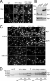
Ypi1 is a nuclear protein. (A) Indirect immunofluorescence with anti-Myc antibody of YPI1–13Myc (JB385-1A) and YPI1-13Myc glc7-129 (JB409-1C) strains. An untagged WT strain (KT1112) serves as the negative control. (B) Immunoblot analysis with anti-Myc antibody of extracts from the YPI1-13Myc (JB385-1A) and GLC7-13Myc (JB412-1B) strains grown to midlog phase in YPD at 30°C. (C) Indirect immunofluorescence of Ypi1-13Myc in WT (JB385-1A) and sds22-6 (JB403-5C) strains at the permissive (24°C) and nonpermissive (37°C) temperatures. (D) Immunoblot analysis of Ypi1-13Myc with anti-Myc antibody of extracts from the strains described in C.
Figure 6.
Figure 6.
Interaction with Ypi1 via the VXW motif is necessary for nuclear targeting and activity of Glc7 in vivo. (A) Diagram of Ypi1 variants and their phenotypes, expressed either from a CEN plasmid or by ectopic integration into the genome. (B) Subcellular distribution of Glc7-mYFP in a ypi1Δ strain carrying either pYPI1 (JB300-3A) or pypi1W53A (JB278-16B). Bar, 5 μm. (C) Subcellular distribution of Sds22-mYFP in a ypi1Δ strain carrying either pYPI1 (JB377-6C) or pypi1W53A (JB378-7D). White arrows indicate nuclei. Bar, 5 μm. (D) Flow cytometry analysis of ypi1Δ mutant cultures containing either pYPI1 (JB300-3A) or pypi1W53A (JB278-16B). Transformants were grown in synthetic medium to midlog phase and processed as described in Materials and Methods. (E) GFP-Tub1 fluorescence in WT (JB309-8B) and ypi1W53A (JB308-7A) cells. Merged DIC and fluorescence images are shown. (F) Serial dilutions of strains were imaged after incubation for 2 d on YPD at the indicated temperatures. The strains used are WT (JB275-1A), ipl1-2 (JB280-5B), ypi1W53A (JB276-8B), ipl1-2 ypi1W53A (JB279-2C), and glc7-127 ipl1-2 (KT1968).
Figure 7.
Figure 7.
Genetic interactions between GLC7 and YPI1. (A and B) Images of serial dilutions of the strains after incubation for 2 d on YPD at designated temperatures. The strains used are YPI1-GFP (JB282-1B), glc7-127 (JB290-1B), YPI1-GFP glc7-127 (JB290-1D), glc7-132 (JB298-5B), YPI1-GFP glc7-132 (JB298-7A), WT (KT1112), glc7-109 (JB297-2A), YPI1-GFP glc7-109 (JB297-1A). The last image in B is that of strains grown on synthetic complete medium that were stained with iodine to assay glycogen levels. Dark color indicates high glycogen levels.

Similar articles

Cited by

References

    1. Alessi D. R., Street A. J., Cohen P., Cohen P. T. Inhibitor-2 functions like a chaperone to fold three expressed isoforms of mammalian protein phosphatase-1 into a conformation with the specificity and regulatory properties of the native enzyme. Eur. J. Biochem. 1993;213:1055–1066. - PubMed
    1. Andrews P. D., Stark M. J. Type 1 protein phosphatase is required for maintenance of cell wall integrity, morphogenesis and cell cycle progression in Saccharomyces cerevisiae. J. Cell Sci. 2000;113:507–520. - PubMed
    1. Axton J. M., Dombradi V., Cohen P.T.W., Glover D. M. One of the protein phosphatase 1 isoenzymes in Drosophila is essential for mitosis. Cell. 1990;63:33–46. - PubMed
    1. Baker S. H., Frederick D. L., Bloecher A., Tatchell K. Alanine scanning mutagenesis of protein phosphatase type 1 in the yeast Saccharomyces cerevisiae. Genetics. 1997;145:615–626. - PMC - PubMed
    1. Black S., Andrews P. D., Sneddon A. A., Stark M. J. A regulated MET3-GLC7 gene fusion provides evidence of a mitotic role for Saccharomyces cerevisiae protein phosphatase 1. Yeast. 1995;11:747–759. - PubMed

Publication types

MeSH terms

Substances