. 2008 Jan 22;117(3):388-95.
doi: 10.1161/CIRCULATIONAHA.107.719765. Epub 2008 Jan 2.

Noninvasive in vivo imaging of monocyte trafficking to atherosclerotic lesions

Affiliations

Noninvasive in vivo imaging of monocyte trafficking to atherosclerotic lesions

Moritz F Kircher et al. Circulation. .

Abstract

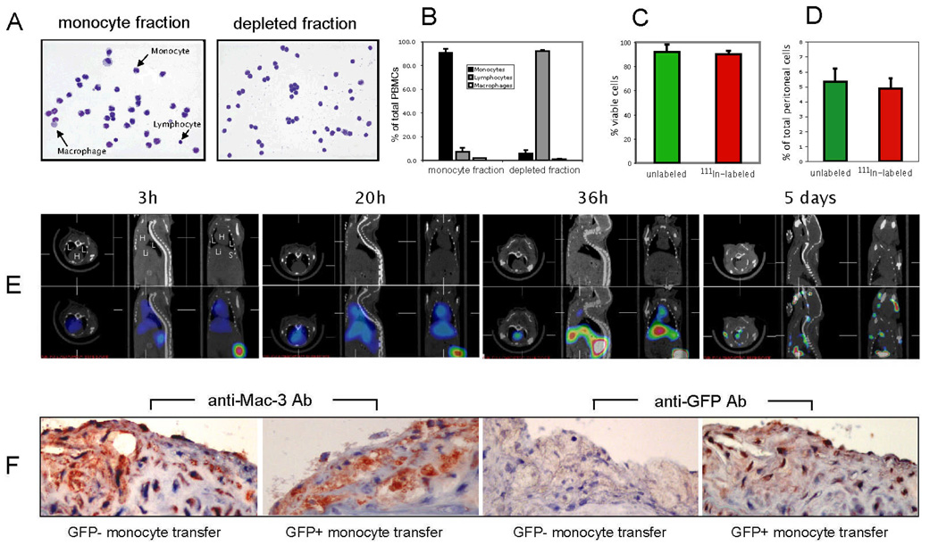
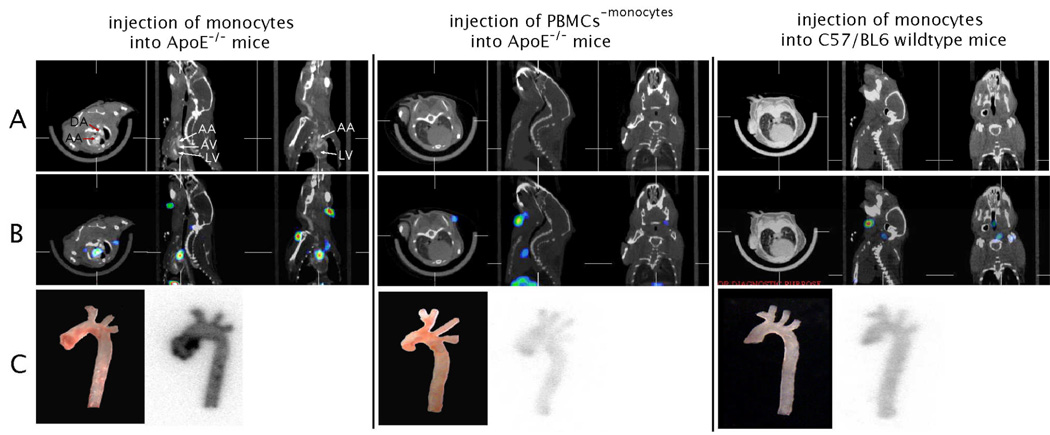
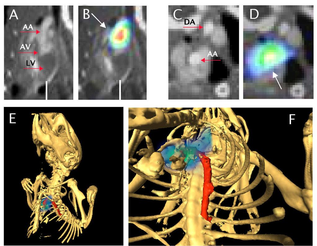
Background: Monocytes play a key role in atherogenesis, but their participation has been discerned largely via ex vivo analyses of atherosclerotic lesions. We sought to establish a noninvasive technique to determine monocyte trafficking to atherosclerotic lesions in live animals.

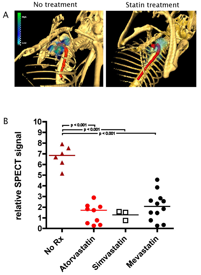
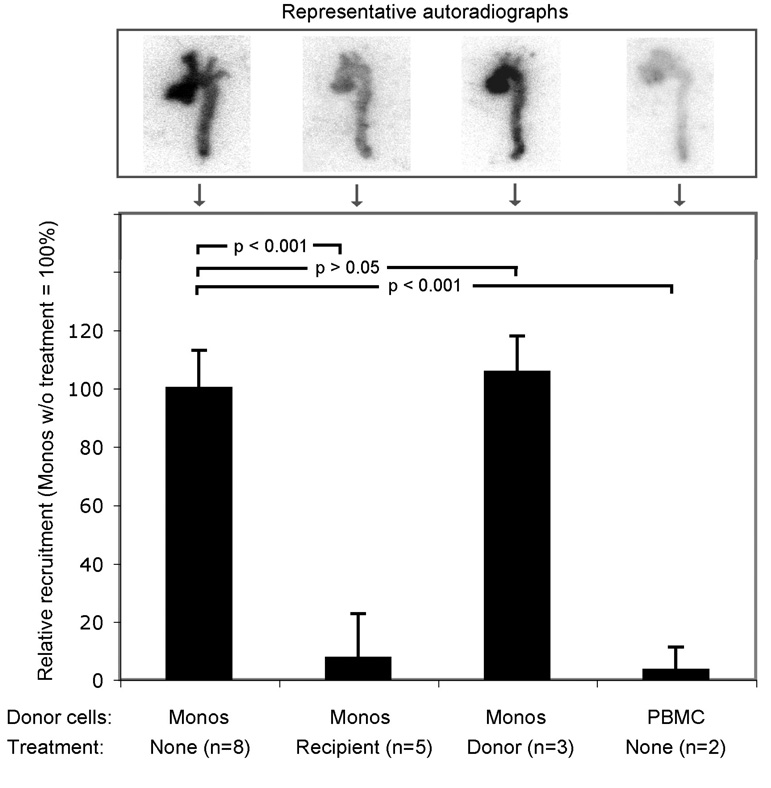
Methods and results: Using a micro-single-photon emission computed tomography small-animal imaging system and a Food and Drug Administration-approved radiotracer ([indium 111] oxyquinoline, (111)In-oxine), we demonstrate here that monocyte recruitment to atherosclerotic lesions can be visualized in a noninvasive, dynamic, and 3-dimensional fashion in live animals. We show in vivo that monocytes are recruited avidly to plaques within days of adoptive transfer. Using micro-single-photon emission computed tomography imaging as a screening tool, we were able to investigate modulatory effects on monocyte recruitment in live animals. We found that 3-hydroxy-3-methylglutaryl coenzyme A reductase inhibitors rapidly and substantially reduce monocyte recruitment to existing atherosclerotic lesions, as imaged here in vivo.

Conclusions: This novel approach to track monocytes to atherosclerotic plaques in vivo should have broad applications and create new insights into the pathogenesis of atherosclerosis and other inflammatory diseases.


Conflict of interest statement

Conflict of Interest Disclosures

None.

Figures

Figure 1
Figure 1. Characterization of monocyte isolation, effect of 111In-oxine labeling on monocyte function, in vivo biodistribution and histological validation
A, Cytospin and Wright-Giemsa staining of monocyte fraction and depleted fraction of PBMCs. B, Quantification of monocyte purity based on manual counting of cytospin slides (summary of three independent experiments). C, Effect of 111In-oxine labeling on monocyte viability as determined by Trypan blue staining. D, Effect of 111In-oxine labeling on the in vivo recruitment capability of monocytes in a model of thioglycollate-induced peritonitis. E, In vivo SPECT/CT imaging time-course of monocyte biodistribution in ApoE−/− mouse (H = heart; L = lung; Li = liver; S = spleen). F, Histological validation of monocyte recruitment to atherosclerotic plaques in ApoE−/− mice 5 days after injection of GFP− or GFP+ monocytes, respectively. Immunohistochemistry using Abs against Mac-3 and GFP (sister sections; positive staining for both in red (note the positive staining in all sections except for anti-GFP staining with transfer of GFP- monocytes). Data are representative of three independent experiments.
Figure 2
Figure 2. microSPECT/CT allows imaging of monocyte recruitment to atherosclerotic plaques
Monocytes vs. PBMCs without monocytes (PBMC-monocytes) were labeled with 111In-oxine and transferred into ApoE−/− mice (left, middle). As a control, monocytes were transferred into C57BL/6 wildtype mice (right). MicroSPECT/CT imaging was performed 5 days after injection of cells. A CT images in axial, sagittal and coronal views, respectively. B, SPECT/CT overlay images in axial, sagittal and coronal views, respectively. C white light images and corresponding autoradiography exposures of aortas excised after in vivo imaging. (LV = left ventricle; AV = aortic valve region; AA = ascending aorta).
Figure 3
Figure 3. Magnification and 3D view of the SPECT/CT data shown in Fig.2 (monocytes transferred into ApoE−/− mouse)
A and C depict CT data only, B and D SPECT/CT overlay. A and B show sagittal views of the heart (LV = left ventricle; AV = aortic valve region) and ascending aorta (AA). Note the SPECT signal (white arrows) indicating monocyte recruitment to the ascending aorta in B. C and D depict axial views of ascending (AA) and descending aorta (DA). Note the SPECT signal in D in the ascending aorta, correlating with the location in B. E and F, 3D rendering; bony structures rendered in white, aorta in red and monocyte recruitment in blue-green scale. See also web-movie for animation of 3D-dataset.
Figure 4
Figure 4. MicroSPECT/CT imaging allows noninvasive assessment of treatment effect of statins drugs on monocyte recruitment
A, Representative example of in vivo imaging of statin treatment. Note the absent activity in the aorta in the animal treated with atorvastatin. Skeleton rendered in white, aorta in red and monocyte recruitment in blue-green scale. B, Differences of in vivo recruitment between placebo-treated animals (no Rx) and animals treated with different statins. Statistics (p-value) for each group were calculated by using one-way ANOVA with Tukey’s multiple-comparison test.
Figure 5
Figure 5. Ex vivo quantification of atorvastatin treatment effect on monocyte recruitment
Adoptive transfer experiments were performed either without treatment or with atorvastatin treatment of recipient or donor mice, respectively. Excised aortas were exposed on autoradiography plates and quantitative analysis of radioactivity performed. Results were expressed as relative recruitment of cells (monocytes without treatment scaled to 100%). Statistics (p-value) for each group were calculated by using one-way ANOVA with Tukey’s multiple-comparison test.

References

    1. Li AC, Glass CK. The macrophage foam cell as a target for therapeutic intervention. Nat Med. 2002;8:1235–1242. - PubMed
    1. Libby P. Inflammation in atherosclerosis. Nature. 2002;420:868–874. - PubMed
    1. Binder CJ, Chang MK, Shaw PX, Miller YI, Hartvigsen K, Dewan A, Witztum JL. Innate and acquired immunity in atherogenesis. Nat Med. 2002;8:1218–1226. - PubMed
    1. Libby P, Ridker PM. Inflammation and atherosclerosis: role of C-reactive protein in risk assessment. Am J Med. 2004;116(Suppl 6A):9S–16S. - PubMed
    1. Ross R. Atherosclerosis--an inflammatory disease. N Engl J Med. 1999;340:115–126. - PubMed

Publication types