. 2008 Jan;49(1):34-41.
doi: 10.1167/iovs.07-0685.

Small proline-rich protein 1B (SPRR1B) is a biomarker for squamous metaplasia in dry eye disease

Affiliations

Small proline-rich protein 1B (SPRR1B) is a biomarker for squamous metaplasia in dry eye disease

Shimin Li et al. Invest Ophthalmol Vis Sci. 2008 Jan.

Abstract

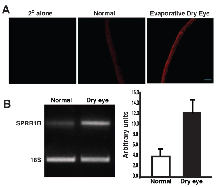
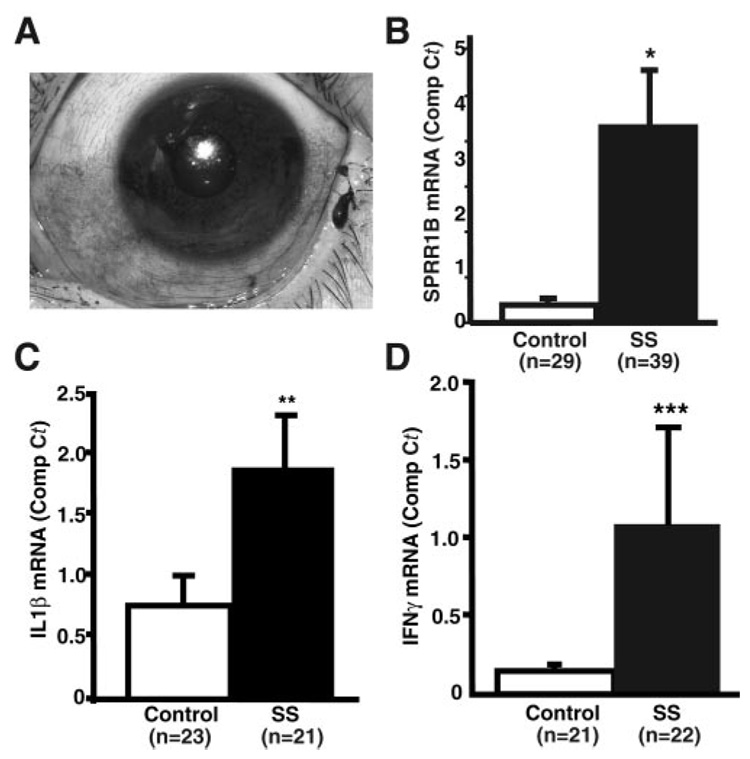
Purpose: Squamous metaplasia occurs in ocular surface diseases like Sjögren's syndrome (SS). It is a phenotypic change whereby epithelial cells initiate synthesis of squamous cell-specific proteins such as small proline-rich protein 1B (SPRR1B) that result in pathologic keratin formation on the ocular surface. The authors hypothesized that inflammation is a key inducer of pathologic keratinization and that SPRR1B represents an analytical biomarker for the study of the molecular mechanisms.

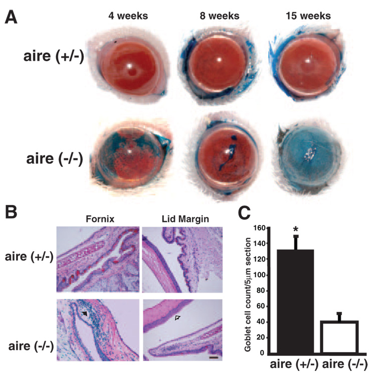
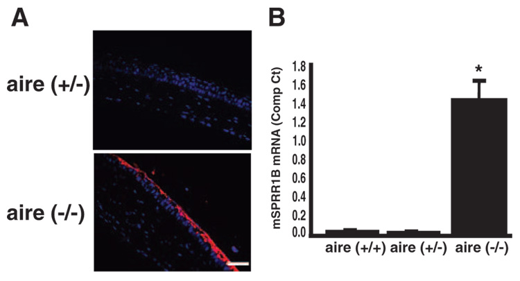
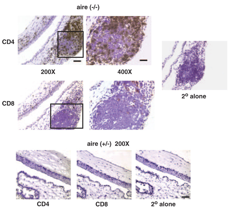
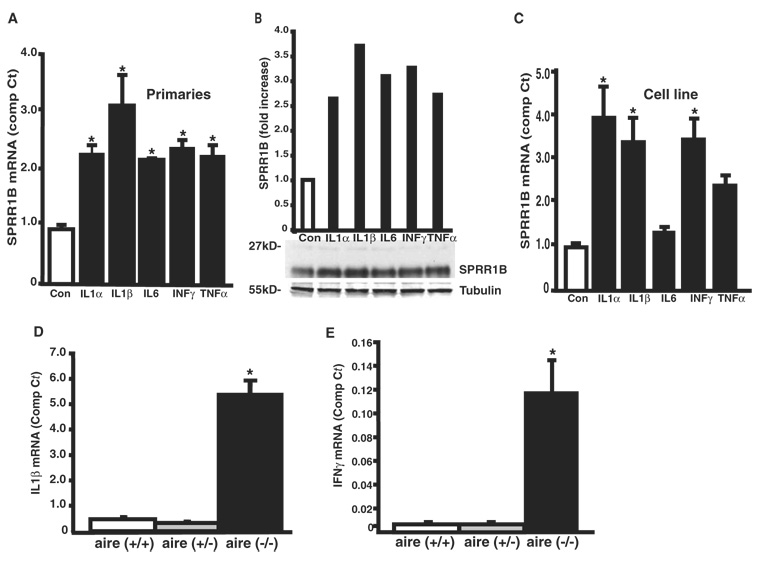
Methods: Real-time quantitative RT-PCR and immunohistochemistry were used to examine SPRR1B mRNA and protein in two different mouse models of dry eye and patients with SS. Adoptive transfer of mature lymphocytes from mice lacking the autoimmune regulator (aire) gene was performed to examine the role of inflammation as an inducer of squamous metaplasia. SPRR1B expression in response to several cytokines was examined in vitro, whereas the expression of cytokines IL1beta and IFNgamma was quantified in ocular tissues of aire-deficient mice and patients with SS.

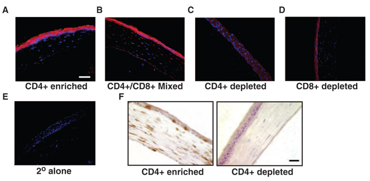
Results: SPRR1B was increased across the ocular surface of mice with both desiccating stress and autoimmune-mediated, aqueous-deficient dry eye and in patients with SS. Adoptive transfer of CD4(+) T cells from aire-deficient mice to immunodeficient recipients caused advanced ocular surface keratinization. IL1alpha, IL1beta, IL6, IFNgamma, and TNFalpha induced SPRR1B expression in vitro and the local expression of IL1beta and IFNgamma was elevated in ocular tissues of patients with SS and aire-deficient mice.

Conclusions: SPRR1B is a valid biomarker for the study of the molecular mechanisms of squamous metaplasia. There is a definitive link between inflammation and squamous metaplasia in autoimmune-mediated dry eye disease, with IL1beta and IFNgamma likely acting as key participants.


Figures

FIGURE 1
FIGURE 1
Corneal epithelial expression of SPRR1B protein and mRNA in an aqueous-deficient– evaporative mouse model of dry eye. (A) Anti-SPRR1B antibody detected with anti-rabbit Cy3 (red) showed increased expression of SPRR1B in a dry-eye mouse (right) compared with a normal one (middle). Secondary antibody alone was negative (left), as was the rabbit IgG isotype control (not shown). SPRR1B-positive immunostaining is representative of that observed in three sections obtained from three different dry eye and control mice. Scale bar, 50 µm. (B) SPRR1B mRNA expression is increased in the desiccating stress mouse model of dry eye. Corneal epithelial cells were scraped from the ocular surface and lysed for RNA extraction after 7 days of cholinergic blockade and air current desiccation. Results were compared to cells obtained from normal control mice (scraped cells were pooled from multiple dry eye and control eyes to generate 1 µg of RNA for each group) and were normalized using primers for 18S RNA. Right: densitometric analysis.
FIGURE 2
FIGURE 2
Dry eye phenotype in aire-deficient mice. (A) Punctate corneal epithelial erosions were apparent with lissamine green staining as early as 4 weeks and appeared to progress to a filamentary keratitis by 8 weeks of age. Moderate to severe disruption of ocular surface integrity was present in all aire−/− mice by 15 weeks. (B) Hallmark histologic signs of dry eye in aire-deficient mice were accompanied by increased epithelial stratification (open black arrow), cellular infiltration of the tarsal conjunctiva and cornea (solid black arrow), and loss of goblet cells. Scale bar, 50 µm; magnification, ×200. (C) Goblet cells were quantified in aire-deficient vs. aire-sufficient mice at 15 weeks by PAS staining. Counts are expressed as mean ± SE per 5-µm section (aire+/− vs. aire−/−, 133 ± 16.0 vs. 45 ± 10.4, respectively; *P < 0.0001).
FIGURE 3
FIGURE 3
The squamous cell biomarker, SPRR1B, was increased across the ocular surface of aire-deficient mice. (A) Representative example of SPRR1B protein expression on the corneal surface of aire-deficient mice at 15 weeks. Anti-SPRR1B antibody detected with Cy3 secondary (red) was associated with the superficial layers of the corneal epithelium in aire−/− vs. littermate control (aire+/−). Magnification, ×400. Blue: nuclei stained with DAPI. Scale bar, 50 µm. (B) Comparison of SPRR1B mRNA expression in the corneas of aire+/+ (wild-type), aire+/− (heterozygous), and aire−/− (knockout) mice using RT-qPCR. A minimum of three eyes from three different animals were combined for each experiment. Experiments were repeated a minimum of three times, and results are expressed as the mean ± SE (wild-type undetected; heterozygous, undetected; knockout = 1.43 ± 0.224; *P < 0.01).
FIGURE 4
FIGURE 4
The ocular surface and lids (box, ×400) of aire-deficient mice are infiltrated with CD4+ and CD8+ T cells. OCT embedded sections of aire−/− BALB/c mice were stained with antibodies directed against CD4+ (top) and CD8+ (middle) T cells, as described. Staining of heterozygous littermates (bottom) was negative for both cell populations. Secondary antibody–alone control staining was negative in all tissues. Specimens are representative of CD4+ and CD8+ immunostaining observed in tissue sections obtained from 8 knockout and 12 heterozygous mice. Scale bar for ×400, 30 µm; for ×200, 50 µm.
FIGURE 5
FIGURE 5
The squamous phenotype can be induced by adoptive transfer of lymphocytes from the regional lymph node and spleen of aire-deficient mice. Pooled lymphocyte populations were (A) enriched for CD4+ T cell, (B) enriched for CD4+ and CD8+ T cells, (C) depleted of CD4+ T cells, or (D) depleted of CD8+ T cells. Cells were transferred via intravenous tail vein injection into immunodeficient (SCID) recipients at 1 × 10⁷ cells per mouse (n = 5 per group). Immunofluorescence with anti-SPRR1B antibody and Cy3 secondary (red) shows localization throughout the corneal epithelium that is absent in eyes of (E) secondary alone control animals and those (C) depleted of CD4+ T cells. (F) Immunohistochemical analysis of the same tissues demonstrated extensive infiltration of the ocular surface with CD4+ T cells (labeled brown with DAB) in CD4+ enriched animals (left) that was absent in CD4+ depleted animals (right). Blue: nuclei stained with DAPI. Magnification, ×400; scale bar, 50 µm.
FIGURE 6
FIGURE 6
Upregulation of SPRR1B (A) mRNA and (B) protein in primary human corneal epithelial cells after a 12-hour exposure to the cytokines IL1α, IL1β, IL6, IFNγ, and TNFα. Real-time PCR results are expressed as the mean ± SE comparative Ct calculated using GAPDH as an endogenous reference (control = 0.92 ± 0.082, IL1α 2.18 ± 0.147, IL1β = 3.00 ± 0.504, IL6 = 2.08 ± 0.026, IFNγ = 2.28 ± 0.132, and TNFα = 2.15 ± 0.183). (C) Similar levels of induction were observed in SV40-HCE cells (control = 0.98 ± 0.095, IL1α = 3.92 ± 0.714, IL1β = 3.37 ± 0.561, IL6 = 1.34 ± 0.114, IFNγ = 3.44 ± 0.475, and TNFα = 2.39 ± 0.237, respectively). (D) IL1β and (E) IFNγ mRNA were significantly increased in aire-deficient mice compared to aire-sufficient littermates. The mean ± SE comparative Ct for IL1β and IFNγ was compared in corneal tissues of wild-type, heterozygous, and knockout mice by RT-qPCR (IL1β = 0.424 ± 0.161, 0.284 ± 0.096, and 5.37 ± 0.878, respectively; IFNγ = undetected, undetected, and 0.120 ± 0.023, respectively) *P < 0.05. Experiments were repeated a minimum of three times. Both IL1β and IFNγ expression correlated highly with SPRR1B expression in aire-deficient mice (r = 0.92, P < 0.001 for IL1β and r = 0.81, P < 0.01 for IFNγ).
FIGURE 7
FIGURE 7
SPRR1B is upregulated in patients with SS. (A) Impression cytology specimens were collected from lissamine green–stained areas of the temporal bulbar conjunctiva in patients with SS and compared to age-matched control subjects without staining. RT-qPCR was used to determine the comparative Ct for (B) SPRR1B, (C) IL1β, and (D) IFNγ. Expressed as the mean ± SE in control versus patients with SS: SPRR1B = 0.31 ± 0.087 vs. 3.37 ± 1.00, *P < 0.001; IL1β = 0.766 ± 0.198 vs. 1.88 ± 0.633, **P = 0.085; and IFNγ = 0.149 ± 0.034 vs. 1.097 ± 0.593, ***P = 0.11.

References

    1. Nakamura T, Nishida K, Dota A, Kinoshita S. Changes in conjunctival clusterin expression in severe ocular surface disease. Invest Ophthalmol Vis Sci. 2002;43:1702–1707. - PubMed
    1. Tsubota K, Satake Y, Kaido M, et al. Treatment of severe ocular-surface disorders with corneal epithelial stem-cell transplantation. N Engl J Med. 1999;340:1697–1703. - PubMed
    1. Sacks EH, Jakobiec FA, Wieczorek R, Donnenfeld E, Perry H, Knowles DM., Jr Immunophenotypic analysis of the inflammatory infiltrate in ocular cicatricial pemphigoid: further evidence for a T cell-mediated disease. Ophthalmology. 1989;96:236–243. - PubMed
    1. Raphael M, Bellefqih S, Piette JC, Le Hoang P, Debre P, Chomette G. Conjunctival biopsy in Sjögren’s syndrome: correlations between histological and immunohistochemical features. Histopathology. 1988;13:191–202. - PubMed
    1. Pflugfelder SC, Huang AJ, Feuer W, Chuchovski PT, Pereira IC, Tseng SC. Conjunctival cytologic features of primary Sjögren’s syndrome. Ophthalmology. 1990;97:985–991. - PubMed

Publication types

MeSH terms