

Activated satellite cells in medial rectus muscles of patients with strabismus
- PMID: 18172095
- PMCID: PMC3039278
- DOI: 10.1167/iovs.07-0507
Activated satellite cells in medial rectus muscles of patients with strabismus
Abstract
Purpose: The goal of this study was to determine whether the medial rectus muscles of patients with a history of medial rectus underaction or overaction show alterations in the process of satellite cell activation when compared with normal age-matched control muscles.
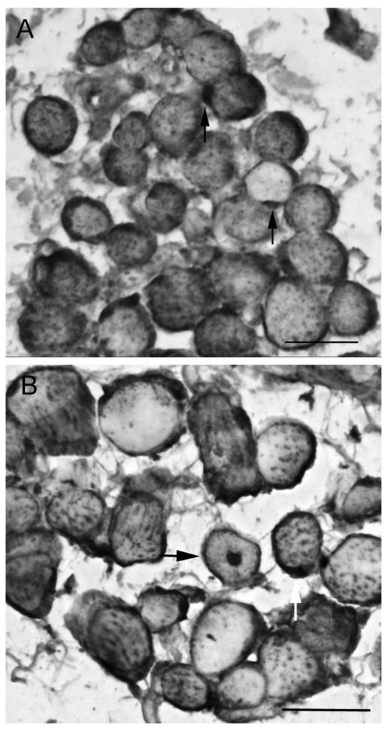
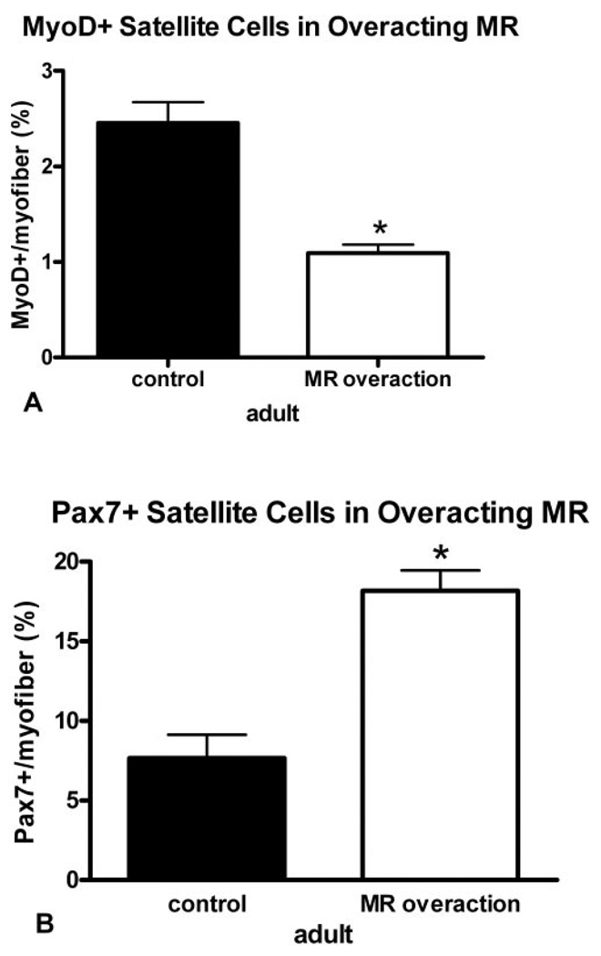
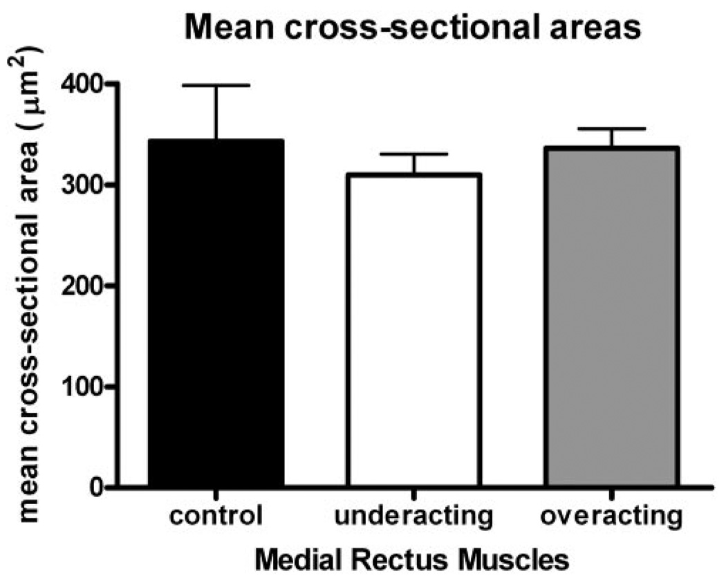
Methods: Medial rectus muscles were obtained with consent from adult patients undergoing surgical resection due to medial rectus underaction or overaction and were prepared for histologic examination by fixation and paraffin embedding. Control muscles were obtained from cornea donor eyes of adults who had no history of strabismus or neuromuscular disease. Cross sections were obtained and stained immunohistochemically for the presence of activated satellite cells, as identified by MyoD immunoreactivity, and the presence of the total satellite cell population, as identified by Pax7 immunoreactivity. The percentages of MyoD- and Pax7-positive satellite cells per 100 myofibers in cross section were calculated.
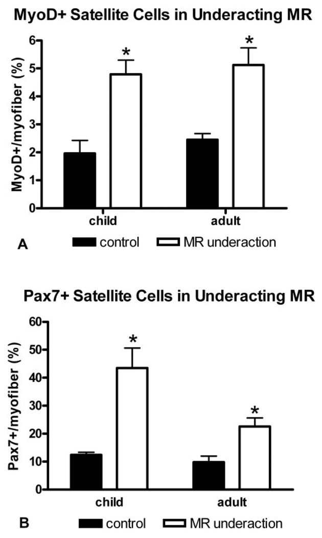
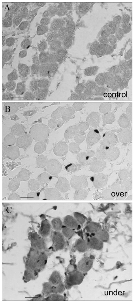
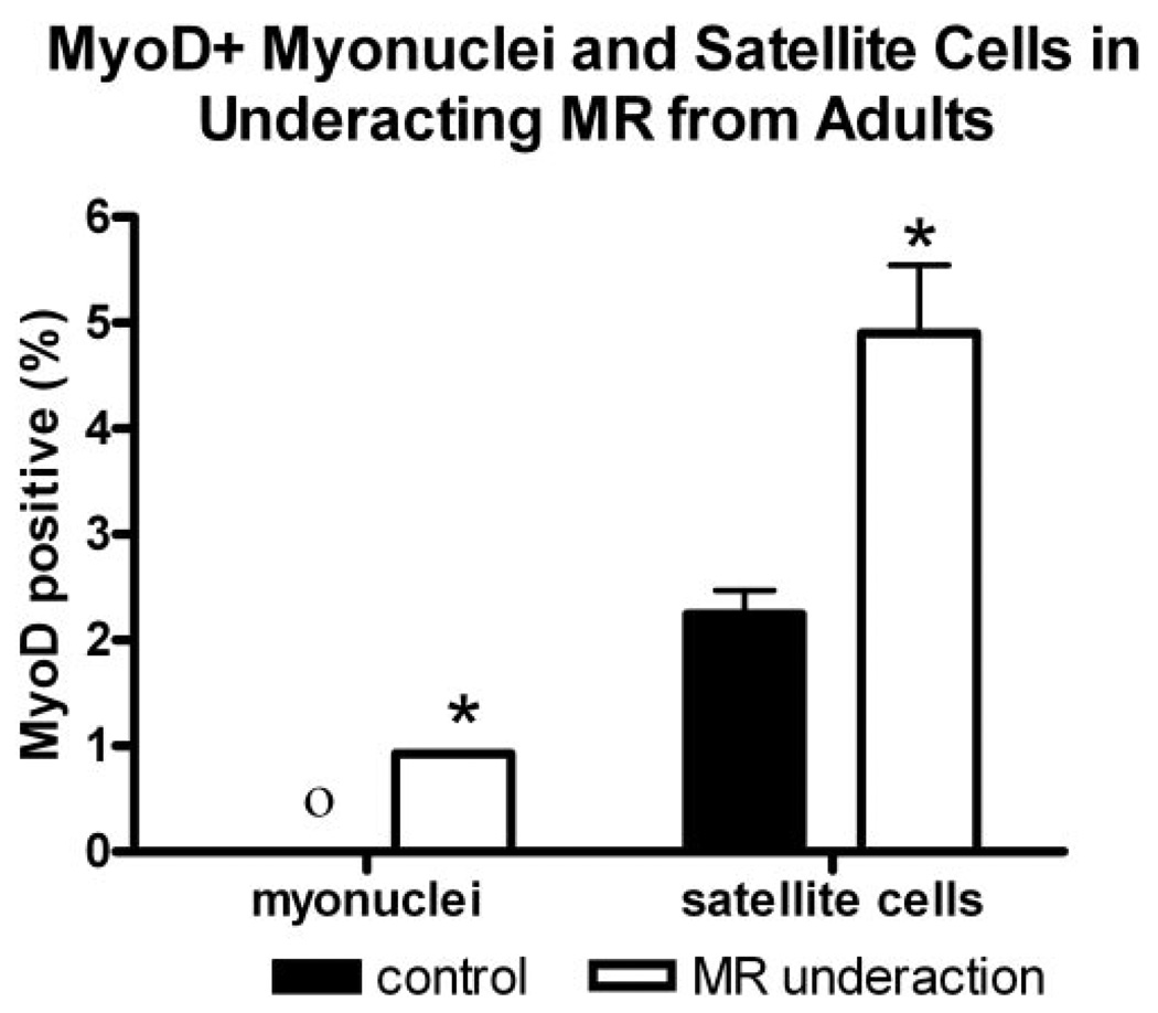
Results: As predicted from results in the literature, MyoD-positive satellite cells, indicative of activation, were present in both the control and resected muscles. In the underacting medial rectus muscles, the percentages of MyoD- and Pax7-positive satellite cells, based on the number of myofibers, were approximately twofold higher than the percentages in the control muscles. In the overacting medial rectus muscles, the percentage of MyoD-positive satellite cells was twofold less than in the control muscles, whereas the percentage of Pax7-positive satellite cells significantly increased compared with that in the control specimens.
Conclusions: The presence of an increased number of activated satellite cells in the resected underacting medial rectus muscles and the decreased numbers of activated satellite cells in the overacting muscles was unexpected. The upregulation in the number of MyoD-positive satellite cells in underacting muscles suggests that there is potential for successful upregulation of size in these muscles, as the cellular machinery for muscle repair and regeneration, the satellite cells, is retained and active in patients with medial rectus underaction. The decreased number of activated satellite cells in overacting MR muscle suggests that factors as yet unknown in these overacting muscles are able to affect the number of satellite cells and/or their responsiveness compared with normal age-matched control muscles. These hypotheses are currently being tested.
Figures
Similar articles
-
Increased frequency of activated satellite cells in overacting inferior oblique muscles from humans.Invest Ophthalmol Vis Sci. 2006 Aug;47(8):3360-5. doi: 10.1167/iovs.05-0798. Invest Ophthalmol Vis Sci. 2006. PMID: 16877403
-
Changes in PAX7 Positive Satellite Cells in Extraocular Muscle After Strabismus Surgery.J Pediatr Ophthalmol Strabismus. 2025 May-Jun;62(3):190-195. doi: 10.3928/01913913-20241210-02. Epub 2025 Jan 21. J Pediatr Ophthalmol Strabismus. 2025. PMID: 39835589
-
Increased myofiber size and reduced satellite cell numbers in medial rectus muscle of patients with intermittent exotropia.Strabismus. 2020 Dec;28(4):201-207. doi: 10.1080/09273972.2020.1832546. Epub 2020 Oct 21. Strabismus. 2020. PMID: 33085552
-
Activated satellite cells in extraocular muscles of normal adult monkeys and humans.Invest Ophthalmol Vis Sci. 2003 May;44(5):1927-32. doi: 10.1167/iovs.02-0673. Invest Ophthalmol Vis Sci. 2003. PMID: 12714625 Free PMC article.
-
The molecular regulation of muscle stem cell function.Cold Spring Harb Symp Quant Biol. 2008;73:323-31. doi: 10.1101/sqb.2008.73.064. Epub 2009 Mar 27. Cold Spring Harb Symp Quant Biol. 2008. PMID: 19329572 Review.
Cited by
-
Childhood Onset Strabismus: A Neurotrophic Factor Hypothesis.J Binocul Vis Ocul Motil. 2021 Apr-Jun;71(2):35-40. doi: 10.1080/2576117X.2021.1893585. Epub 2021 Apr 19. J Binocul Vis Ocul Motil. 2021. PMID: 33872122 Free PMC article.
-
Effects of recession versus tenotomy surgery without recession in adult rabbit extraocular muscle.Invest Ophthalmol Vis Sci. 2010 Nov;51(11):5646-56. doi: 10.1167/iovs.10-5523. Epub 2010 Jun 10. Invest Ophthalmol Vis Sci. 2010. PMID: 20538996 Free PMC article.
-
Morphological Differences in the Inferior Oblique Muscles from Subjects with Over-elevation in Adduction.Invest Ophthalmol Vis Sci. 2020 Jun 3;61(6):33. doi: 10.1167/iovs.61.6.33. Invest Ophthalmol Vis Sci. 2020. PMID: 32539136 Free PMC article.
-
A Preliminary Study of the Occurrence of Genetic Changes in mtDNA in the Muscles in Children Treated for Strabismus.J Clin Med. 2024 Jul 10;13(14):4041. doi: 10.3390/jcm13144041. J Clin Med. 2024. PMID: 39064081 Free PMC article.
-
Differences in gene expression between strabismic and normal human extraocular muscles.Invest Ophthalmol Vis Sci. 2012 Aug 3;53(9):5168-77. doi: 10.1167/iovs.12-9785. Invest Ophthalmol Vis Sci. 2012. PMID: 22786898 Free PMC article.
References
-
- Guyton DL. The 10th Bielschowsky Lecture. Changes in strabismus over time: the role of vergence tonus and muscle length adaptation. Binocul Vis Strabismus Q. 2006;21:81–92. - PubMed
-
- Kushner BJ. Perspective on strabismus. Arch Ophthalmol. 2006;124:1321–1326. - PubMed
-
- Livir-Rallatos G, Gunton KB, Calhoun JH. Surgical results in large-angle exotropia. J AAPOS. 2002;6:77–80. - PubMed
-
- Triger L, Siatkowski RM. Factors associated with horizontal reoperation in infantile esotropia. J AAPOS. 2002;6:15–20. - PubMed
-
- Yamada K, Andrews C, Chan WM, et al. Heterozygous mutations of the kinesin KIF21A in congenital fibrosis of the extraocular muscles type 1 (CFEOM1) Nat Genet. 2003;35:318–321. - PubMed
Publication types
MeSH terms
Substances
Grants and funding
LinkOut - more resources
Full Text Sources
Research Materials
