. 2008 Jan 1;22(1):50-65.
doi: 10.1101/gad.1622708.

Degradation of histone mRNA requires oligouridylation followed by decapping and simultaneous degradation of the mRNA both 5' to 3' and 3' to 5'

Affiliations

Degradation of histone mRNA requires oligouridylation followed by decapping and simultaneous degradation of the mRNA both 5' to 3' and 3' to 5'

Thomas E Mullen et al. Genes Dev. .

Abstract

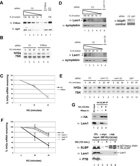
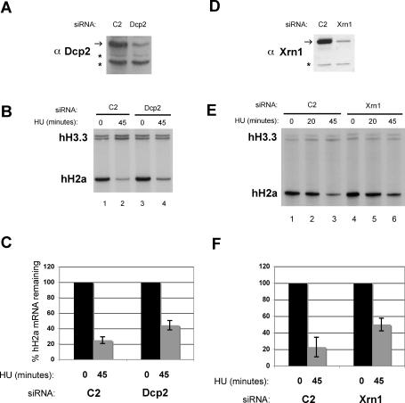
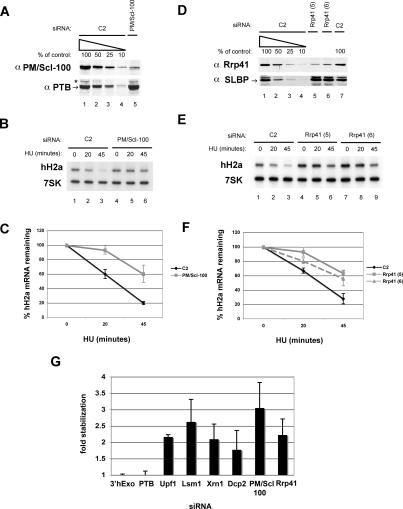
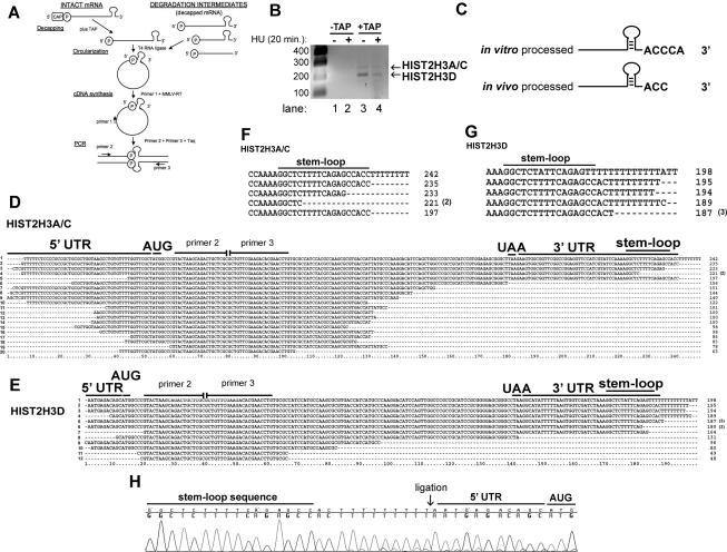
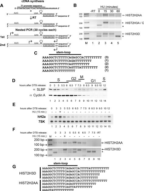
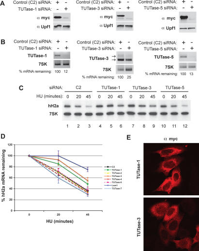
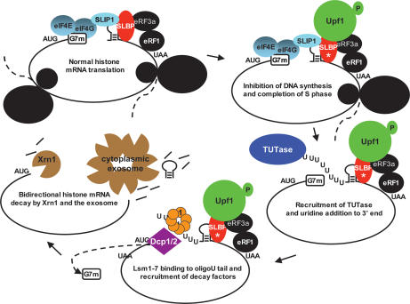
Histone mRNAs are rapidly degraded at the end of S phase or when DNA replication is inhibited. Histone mRNAs end in a conserved stem-loop rather than a poly(A) tail. Degradation of histone mRNAs requires the stem-loop sequence, which binds the stem-loop-binding protein (SLBP), active translation of the histone mRNA, and the location of the stem-loop close to the termination codon. We report that the initial step in histone mRNA degradation is the addition of uridines to the 3' end of the histone mRNA, both after inhibition of DNA replication and at the end of S phase. Lsm1 is required for histone mRNA degradation and is present in a complex containing SLBP on the 3' end of histone mRNA after inhibition of DNA replication. We cloned degradation intermediates that had been partially degraded from both the 5' and the 3' ends. RNAi experiments demonstrate that both the exosome and 5'-to-3' decay pathway components are required for degradation, and individual histone mRNAs are then degraded simultaneously 5' to 3' and 3' to 5'.


Figures

Figure 1.
Figure 1.
Effect of knockdown of 3′hExo or Lsm1 on histone mRNA degradation. HeLa cells were treated with the 3′hExo (AC) or Lsm1 (DF) siRNAs as described in Materials and Methods, and then treated with 5 mM HU and total cell RNA prepared 0, 20, or 45 min after HU treatment. (A,D) A dilution series of the control (C2) cell lysate together with a lysate from cells treated with the indicated siRNA was analyzed by Western blotting for 3′hExo and Upf1. The asterisk indicates a cross-reacting band. (B,E) Two micrograms of total cell RNA were resolved by urea acrylamide gel electrophoresis, transferred to N+ nitrocellulose, and probed with a mixture of histone H2a mRNA and 7SK snRNA probes. (C,F) The average of three independent experiments is shown. (C) (◆) C2; (■) 3′hExo. (F) (◆) C2; (■) Lsm1-1; (▴) Lsm1-2; (●) Upf1. Standard deviations are indicated by vertical bars. (G) Lysates were prepared from S-phase cells expressing HA-SLBP 15 min after treatment with HU as described previously (Kaygun and Marzluff 2005a). (Top, lane 3) A portion of the lysate was treated with RNase A (20 μg/mL) for 15 min prior to immunoprecipitation with anti-HA antibody. The immunoprecipitates were analyzed for SLBP and Lsm1 by Western blotting. (Bottom) S-phase cells expressing HA-SLBP were treated with HU for 15 min; lysates were prepared and subjected to immunoprecipitation with anti-myc (lanes 3,4) or anti-HA (lanes 5,6) antibodies. The immunoprecipitates were analyzed for SLBP, Lsm1, and PTB as a control by Western blotting. (Lanes 1,2) The input lanes show 5% of the total lysate.
Figure 2.
Figure 2.
Effect of decapping complex and 5′–3′ exonuclease knockdown on histone mRNA degradation. HeLa cells were treated with the indicated siRNAs to Dcp2 (AC) or Xrn1 (DF) and treated with 5 mM HU as described in Figure 1. (A,D) Protein levels of knockdowns were measured by Western blot using antibodies to Dcp2 (A) and Xrn1 (D). (B,E) S1 nuclease protection assays of 5 μg of total RNA isolated from cells treated with 5 mM Hu for 0, 20, or 45 min from Dcp2 (B) or Xrn1 (E) knockdowns. Human H3.3 and H2a mRNAs were detected by mixing probes to detect the 3′ and 5′ ends of H3.3 and H2a mRNAs, respectively. (C,F) The average of three independent experiments from Dcp2 (C) and Xrn1 (F) knockdowns is shown. The vertical bars indicate the standard deviation.
Figure 3.
Figure 3.
Effect of knockdown of exosome components on histone mRNA degradation. (A,D) HeLa cells were treated with the indicated siRNAs to PM/Scl-100 (AC) or Rrp41 (DF) and were treated with 5 mM HU, and the levels of each protein were determined by Western blotting as in Figures 1 and 2. (B,E) Two micrograms of total cell RNA from cells with PM/Scl100 knocked down (B) or Rrp41 knocked down with two siRNAs (5 and 6) to Rrp41 were resolved by urea-acrylamide gel electrophoresis and probed with a mixture of histone H2a mRNA and 7SK snRNA probes as in Figure 1. (C,F) The average of three independent knockdowns to PM/Scl-100 (C: ◆, C2; ■, PM/Scl-100) and Rrp41 [F: ◆, C2; ■, Rrp41(1); -▴-, Rrp41(2)] is shown. (G) Average fold stabilization of all experiments shown in Figures 1, 2, 3, with standard deviations indicated by vertical bars.
Figure 4.
Figure 4.
Detection of the 5′ and 3′ ends of capped histone H3 mRNA in vivo and determination of the sequence of decapped histone H3 mRNA degradation intermediates. (A) The cRT–PCR strategy to determine the ends of intact histone mRNA and degradation intermediates is shown (adapted from Couttet et al. 1997; © 1997 National Academy of Sciences, USA). (B) cRT–PCR reactions with and without decapping from total cell RNA were separated on 1.5% agarose gels, and amplicons were detected by ethidium bromide staining. Two human histone H3 mRNAs are visualized in the +TAP lanes. (C) The 3′ end of a synthetic histone pre-mRNA substrate processed in vitro and decapped differs from histone mRNA decapped and retrieved from the cell. In vitro processed pre-mRNA and total cell RNA were decapped (+TAP), subjected to cRT–PCR, and cloned. Complete sequences of these experiments are presented in Supplemental Figure S4. (D,E) cRT–PCR was performed on total cell RNA, and the histone H3 products were amplified, cloned, and sequenced. HIST2H3A/C (D) or HIST2H3D (E) mRNAs that were not capped were cloned by cRT–PCR. Primers 2 and 3 represent sites where we targeted amplification toward the 5′ and 3′ ends. ORF sequences between these two oligonucleotides are missing. Numbers in parentheses are the number of times each clone was obtained. (F,G) The 3′ ends from the largest HIST2H3A/C (F) and HIST2H3D (G) mRNA degradation intermediates are shown. The number of times each sequence was obtained is indicated by a number in parentheses. (H) Chromatogram from a HIST2H3D mRNA degradation intermediate containing eight untemplated Us (Ts in the DNA sequence). The 3′ and 5′ ends were ligated (arrow) together.
Figure 5.
Figure 5.
Detection of oligouridylated histone mRNAs after inhibition of DNA synthesis and at the end of S phase. (A) RT–PCR strategy to detect oligo(U)-containing histone mRNA molecules. Total RNA from HeLa cells was primed for cDNA synthesis using an oligo(dA) primer fused to a T7 sequence. Two rounds of PCR (30 cycles each) were performed using oligos targeting the 5′ UTR (first round) or the ORF (second round) and a primer complementary to the T7 sequence. (B) Oligo(dA) RT–PCR from HIST2H2AA, HIST2H3A/C, and HIST2H3D treated with HU for 0, 15, 30, and 60 min. The products were resolved by agarose gel electrophoresis and were detected by ethidium bromide staining. Asterisk indicates a nonspecific band. (C) Sequences from oligo(dA) RT–PCR reactions from the HIST2H3D mRNA when treated with HU for 15 min. The number in parentheses indicates the number of times a particular sequence was observed. (D) HeLa cells were synchronized by DTB and released into S phase, and protein samples were collected at the indicated times. Western blot analysis of SLBP and cyclin A was performed. (E) Total RNA was isolated from the same cells as in D, and H2a mRNA and 7SK RNA levels were measured by Northern blotting. (F) Oligo(dA) RT–PCR on total RNA from the middle and end of S phase into the G2 phase of the cell cycle. Total RNA from the indicated time points following release from DTB was subjected to the RT–PCR strategy depicted in A to detect HIST2H2AA and HIST2H3D mRNA and was resolved on a 2.0% agarose gel. HU was added at 3 h for 15 min in lane 3. (G) The oligo(dA) primed RT–PCR products from the 5.5- and 6.0-h time points were cloned, and several clones from the HIST2H2AA and HIST2H3D mRNAs were cloned and sequenced.
Figure 6.
Figure 6.
RNAi screen to seven TUTases present in the human genome identifies two putative TUTase involved in histone mRNA degradation. The seven TUTases were knocked down using specific siRNAs as in Figure 1. (A) A representative experiment to determine the efficacy of TUTase siRNAs. Myc epitope-tagged TUTases were cloned and transfected at the time of the second hit of siRNA (see Materials and Methods). Protein lysates were harvested 48 h following the second hit and probed by Western using an anti-myc antibody. Upf1 was used as a loading control. (B) Endogenous TUTase mRNA levels were measured by RT–PCR. Two micrograms of total RNA were random primed and subjected to MMLV reverse transcriptase followed by PCR. The degree of knockdown, relative to 7SK RNA as a control, estimated by RT–PCR is indicated. (C) Knockdown of TUTase-1 and TUTase-3 stabilizes histone H2a degradation following inhibition of DNA synthesis by HU. Two micrograms of total cell RNA from cells treated with 5 mM HU were resolved by urea acrylamide gel electrophoresis and analyzed with a mixture of histone H2a mRNA and 7SK snRNA probes. (D) The rate of degradation of histone mRNA in three independent experiments where the different TUTases were knocked down. Lsm1 was included as a positive control. Standard deviations are represented by vertical bars. The U6 TUTase is essential for cell viability, and the effect on histone mRNA degradation could not be determined (see text for details). (E) TUTase-1 and TUTase-3 are localized predominantly in the cytoplasm of HeLa cells. Cells were transfected with myc-tagged clones of TUTase-1 or TUTase-3, and the tagged proteins were detected by immunofluorescence 48 h following transfection.
Figure 7.
Figure 7.
Model of histone mRNA degradation. A proposed model of histone mRNA degradation showing the transition of histone mRNA from active translation as a circular mRNA, followed by the recruitment of Upf1 to the 3′ end (Kaygun and Marzluff 2006) when DNA replication is inhibited, followed by oligouridylation and degradation. SLBP is essential for translation (Sanchez and Marzluff 2002), and SLIP1 bridges SLBP to the 5′ end of histone mRNA (Cakmakci et al. 2007).

Comment in

References

    1. Allmang C., Petfalski E., Podtelejnikov A., Mann M., Tollervey D., Mitchell P. The yeast exosome and human PM-Scl are related complexes of 3′ → 5′ exonucleases. Genes & Dev. 1999;13:2148–2158. - PMC - PubMed
    1. Amrani N., Ganesan R., Kervestin S., Mangus D.A., Ghosh S., Jacobson A. A faux 3′-UTR promotes aberrant termination and triggers nonsense-mediated mRNA decay. Nature. 2004;432:112–118. - PubMed
    1. Blum E., Carpousis A.J., Higgins C.F. Polyadenylation promotes degradation of 3′-structured RNA by the Escherichia coli mRNA degradosome in vitro. J. Biol. Chem. 1999;274:4009–4016. - PubMed
    1. Cakmakci N.G., Lerner R.S., Wagner E.J., Zheng L.-X., Marzluff W.F. SLIP1, a factor required for activation of histone mRNA translation by the stem–loop binding protein. Mol. Cell. Biol. 2007 doi: 10.1128/MCB.01500-07. - DOI - PMC - PubMed
    1. Caponigro G., Parker R. Mechanisms and control of mRNA turnover in Saccharomyces cerevisiae. Microbiol. Rev. 1996;60:233–249. - PMC - PubMed

Publication types