. 2008 Jan 1;22(1):79-90.
doi: 10.1101/gad.1601308.

APC/C-Cdh1-mediated degradation of the Polo kinase Cdc5 promotes the return of Cdc14 into the nucleolus

Affiliations

APC/C-Cdh1-mediated degradation of the Polo kinase Cdc5 promotes the return of Cdc14 into the nucleolus

Clara Visintin et al. Genes Dev. .

Abstract

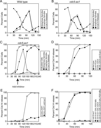
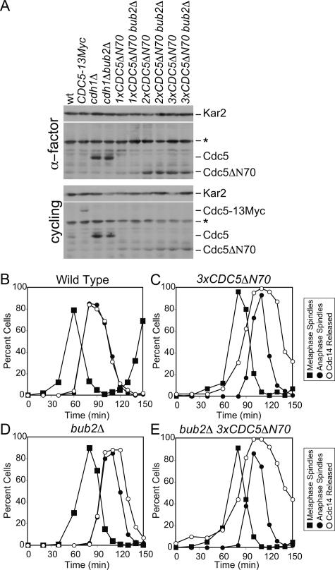
In the budding yeast Saccharomyces cerevisiae, the protein phosphatase Cdc14 triggers exit from mitosis by promoting the inactivation of cyclin-dependent kinases (CDKs). Cdc14's activity is controlled by Cfi1/Net1, which holds and inhibits the phosphatase in the nucleolus from G1 until metaphase. During anaphase, two regulatory networks, the Cdc14 Early Anaphase Release (FEAR) network and the Mitotic Exit Network (MEN), promote the dissociation of Cdc14 from its inhibitor, allowing the phosphatase to reach its targets throughout the cell. The molecular circuits that trigger the return of Cdc14 into the nucleolus after the completion of exit from mitosis are not known. Here we show that activation of a ubiquitin ligase known as the Anaphase-Promoting Complex or Cyclosome (APC/C) bound to the specificity factor Cdh1 triggers the degradation of the Polo kinase Cdc5, a key factor in releasing Cdc14 from its inhibitor in the nucleolus.


Figures

Figure 1.
Figure 1.
De novo protein synthesis is not required for Cdc14 return into the nucleolus. cdc15-2 cells carrying a CDC14-3HA fusion (A1674) were arrested in YEPD at the restrictive temperature (37°C) for 2.5 h. Cells were then released at the permissive temperature (25°C) into YEPD in the absence (A) or presence (B) of 1 mg/mL cyclohexymide. Samples were taken at the indicated times to determine the percentage of cells with anaphase spindles (closed circles) and of cells with Cdc14 released from the nucleolus (open circles). Mitotic CDK inactivation was assayed by analyzing Clb2 protein levels and accumulation of Sic1. Note that the absence of Sic1 in the cycloheximide-treated culture indicates that translation was effectively inhibited. Kar2 was used as an internal loading control in Western blots.
Figure 2.
Figure 2.
APC/C-Cdh1-dependent proteolysis is required for the return of Cdc14 in the nucleolus. (AD) Wild-type (A2747), bub2Δ (A5954), cdh1Δ (A5952), and bub2Δ cdh1Δ (A5950) cells carrying a CDC14-3HA fusion were arrested in G1 in YEPD medium with α-factor pheromone (5 μg/mL). When the arrest was complete (after 3 h), cells were released into medium lacking pheromone. The percentage of budded cells (open diamonds), cells with metaphase spindles (closed squares), and anaphase spindles (closed circles), as well as the percentage of cells with Cdc14-HA released from the nucleolus (open circles), was determined at the indicated times. In E, cells with interphase microtubules were selected at the indicated time points to determine the percentage of cells with Cdc14 released from the nucleolus (n = 100).
Figure 3.
Figure 3.
Clb–CDK or Spo12 inactivation is not required for the return of Cdc14 into the nucleolus. (AC) pMET3-CDC20 pds1Δ cdh1Δ pGAL-SIC1 (A13767) cells were grown in medium lacking methionine supplemented with 2% raffinose (−MetR). Cells were transferred in YEP supplemented with 2% raffinose (YEPR) medium containing 8 mM methionine to arrest pMET3-CDC20 pds1Δ cdh1Δ cells in anaphase. Six hours after the transfer, when >70% of cells were budded and had segregated DNA masses (binucleate cells), the culture was split in two. One-half was maintained in the same medium, whereas 2% galactose was added to the other half to induce SIC1 expression. Samples were taken at the indicated times to analyze the percentage of binucleate cells (A) and Clb2 and Sic1 protein levels and Clb2-CDK kinase activity (C). In B, anaphase cells were selected at the indicated time points to determine the percentage of cells that had Cdc14 released from the nucleolus (n = 100 per time point). (DF) pMET3-CDC20 pds1Δ pGAL-URL-3HA-SPO12 (A13304) cells were grown in medium lacking methionine supplemented with 2% raffinose and 0.5% galactose (−MetR0.5%G). Cells were arrested in early S phase with hydroxyurea (HU; 10 mg/mL) for 3 h followed by release into −MetR0.5%G medium containing 8 mM methionine to arrest pMET3-CDC20 pds1Δ cells in anaphase. Three-and-a-half hours after the release, when ∼90% of cells were budded and had segregated DNA masses (binucleate cells), the culture was split in two. One-half was maintained in the same medium, whereas 2% glucose was added to the other half to deplete Spo12. Samples were taken at the indicated times to analyze the percentage of binucleate cells (D) and Spo12 and Clb2 protein levels (F). In E, anaphase cells were selected at the indicated time points to determine the percentage of cells that had Cdc14 released from the nucleolus (n = 100 per time point).
Figure 4.
Figure 4.
Cdc5’s role in promoting Cdc14 release is restricted to metaphase and anaphase. (A,B) Wild-type (A1411; A) and cdc5-as1 (cdc5L158G; Ry1260; B) cells carrying a CDC14-3HA fusion were arrested in G1 in YEPD with α-factor pheromone (5 μg/mL). After 3 h, cells were released into medium lacking pheromone but containing nocodazole (15 μg/mL) and the CMK inhibitor (5 μM). When the arrest was complete, cells were released into YEPD supplemented with 1% DMSO. The percentage of cells with metaphase spindles (closed squares) and anaphase spindles (closed circles), as well as the percentage of cells with Cdc14-HA released from the nucleolus (open circles), was determined at the indicated times. Note: Spindle morphology was not analyzed 0 and 15 min after release from the nocodazole block because the spindle had not yet reformed. (C) cdc5-as1 GAL-ESP1 CDC14-3HA (Ry1287) cells were arrested in G1 in YEP + 2% Raffinose (YEPR) with α-factor pheromone (5 μg/mL). Two-and-a-half hours into the arrest, 2% galactose (YEPR + G) was added to induce GAL-ESP1 expression. When the arrest was complete (3 h total), cells were released into YEPR + G lacking the pheromone. Seventy-five minutes after the release, when ∼80% of cells had formed a bud and were about to enter metaphase, CMK inhibitor (5 μM) was added. The percentage of budded cells (open diamonds), cells with metaphase spindles (closed squares) and anaphase spindles (closed circles), and the percentage of cells with Cdc14-HA released from the nucleolus (open circles) was determined at the indicated times. (D) Wild-type (A1411; closed circles), cdc15-as1 (A11617; open diamonds), GAL-CDC5 (A3317; open circles), and cdc15-as1 GAL-CDC5 (Ry1245; closed diamonds) cells carrying CDC14-3HA were arrested in G1 in YEP supplemented with 2% Raffinose (YEPR) with α-factor pheromone (5 μg/ mL). After 3 h, cells were released into medium lacking pheromone but supplemented with hydroxyurea (10 mg/mL) and the cdc15-as1 inhibitor (5 μM). After 2.5 h, when cells had arrested in S phase, 2% galactose was added to induce the expression of CDC5 from the inducible GAL1-10 promoter. The percentage of cells with Cdc14-HA released from the nucleolus was determined at the indicated times. (E) GAL-ESP1 (A4150) and cdc5-as1 GAL-ESP1 (Ry1287) cells carrying a CDC14-3HA fusion were treated as described in Figure 6C, except that cells were released into YEPR + G lacking pheromone but supplemented with 15 μg/mL nocodazole. (F) cdc15-as1 (Ry1386; open circles), cdc15-as1 GAL-PDS1dBΔ (Ry1385; closed diamonds), cdc15-as1 GAL-CDC5 (Ry1384; closed circles), and cdc15-as1 GAL-PDS1dBΔ GAL-CDC5 (Ry1383; open diamonds) cells were arrested with hydroxyurea (10 mg/mL). When the arrest was complete, the cdc15-as1 inhibitor (5 μM) was added. One hour after the inhibitor addition, 2% galactose was added to induce the expression of CDC5 and PDS1dBΔ from the inducible GAL1-10 promoter. The percentage of cells with Cdc14-HA released from the nucleolus was determined at the indicated times.
Figure 5.
Figure 5.
CDC5 is required for maintaining Cdc14 in its release state in cdh1Δ cells during anaphase. (AC) pMET3-CDC20 pds1Δ pGAL-URL-3HA-CDC5 (A12396) cells were grown in medium lacking methionine supplemented with 2% raffinose and 0.5% galactose (−MetR0.5%G). Cells were arrested in early S phase with hydroxyurea (HU; 10 mg/mL) for 3 h followed by release into −MetR0.5%G medium containing 8 mM methionine to arrest pMET3-CDC20 pds1Δ cells in anaphase. Three hours after the release, when ∼70% of cells were budded and had segregated DNA masses (binucleate cells), the culture was split in two. One-half was maintained in the same medium, whereas 2% glucose was added to the other half to deplete Cdc5. Samples were taken at the indicated times to analyze the percentage of binucleate cells (A) and Cdc5 and Clb2 protein levels (C). In B, anaphase cells were selected at the indicated time points to determine the percentage of cells that had Cdc14 released from the nucleolus (n = 100 per time point). (DF) pMET3-CDC20 pds1Δ pGAL-URL-3HA-CDC5 cdh1Δ (A12595) cells were treated as described in AC with the exception that it took 3.5 h to arrest cells in the cdc20Δ pds1Δ block instead of 3 h. Samples were taken at the indicated times to analyze the percentage of binucleate cells (D) and Cdc5 and Clb2 protein levels (F). In E, anaphase cells were selected at the indicated time points to determine the percentage of cells that had Cdc14 released from the nucleolus (n = 100 per time point). (GI) pMET3-CDC20 pds1Δ pGAL-URL-3HA-CDC5 pGAL-URL-3HA-SPO12 (A13804) cells were treated as described in AC. Samples were taken at the indicated times to analyze the percentage of binucleate cells (G) and Cdc5, Spo12 and Clb2 protein levels (I). In H, anaphase cells were selected at the indicated time points to determine the percentage of cells that had Cdc14 released from the nucleolus (n = 100 per time point).
Figure 6.
Figure 6.
Cdc5 degradation is important for the timely return of Cdc14 into the nucleolus. (A) Wild-type (A2587), CDC5-13MYC (A2976), cdh1Δ (A5952), cdh1Δ bub2Δ (A5950), 1xCDC5ΔN70 (A10203), 1xCDC5ΔN70 bub2Δ (A10202), 2xCDC5ΔN70 (A10206), 2xCDC5ΔN70 bub2Δ (A10356), 3xCDC5ΔN70 (A10853), and 3xCDC5ΔN70 bub2Δ (A10856) were either arrested in G1 in YEPD medium with α-factor (5 μg/mL) or grown to exponential phase. Cdc5 protein levels were assessed by Western blot. Asterisk (*) marks a cross-reacting band on Western blots. The number of CDC5ΔN70 integrants was assessed by Southern blot analysis. (BE) Wild-type cells (A2747; B), cells carrying three copies of CDC5ΔN70 construct (3xCDC5ΔN7; Α10853; C), bub2Δ cells (A5954; D) and bub2Δ 3xCDC5ΔN70 cells (Α10856; Ε) carrying a CDC14-3HA fusion were arrested with α-factor (5 μg/mL). After 3 h, cells were released into medium lacking pheromone, and the percentage of cells with metaphase spindles (closed squares), anaphase spindles (closed circles), and with Cdc14 released from the nucleolus (open circles) was determined at the indicated times.
Figure 7.
Figure 7.
A model for how APC/C-Cdh1 promotes the return of Cdc14 into the nucleolus. See text for details.

References

    1. Amon A., Irniger S., Nasmyth K. Closing the cell cycle circle in yeast: G2 cyclin proteolysis initiated at mitosis persists until the activation of G1 cyclins in the next cycle. Cell. 1994;77:1037–1050. - PubMed
    1. Azzam R., Chen S.L., Shou W., Mah A.S., Alexandru G., Nasmyth K., Annan R.S., Carr S.A., Deshaies R.J. Phosphorylation by cyclin B–Cdk underlies release of mitotic exit activator Cdc14 from the nucleolus. Science. 2004;305:516–519. - PubMed
    1. Bachmair A., Finley D., Varshavsky A. In vivo half-life of a protein is a function of its amino-terminal residue. Science. 1986;234:179–186. - PubMed
    1. Bardin A.J., Amon A. Men and sin: What’s the difference? Nat. Rev. Mol. Cell Biol. 2001;2:815–826. - PubMed
    1. Bishop A.C., Buzko O., Shokat K.M. Magic bullets for protein kinases. Trends Cell Biol. 2001;11:167–172. - PubMed

Publication types

MeSH terms