. 2008 Jan 1;22(1):106-20.
doi: 10.1101/gad.1590908.

Axin and GSK3- control Smad3 protein stability and modulate TGF- signaling

Affiliations

Axin and GSK3- control Smad3 protein stability and modulate TGF- signaling

Xing Guo et al. Genes Dev. .

Abstract

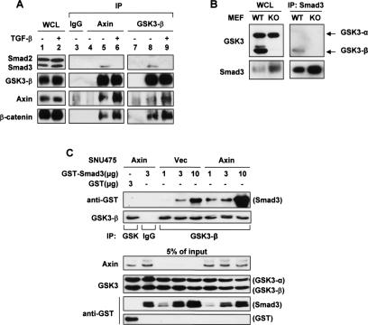
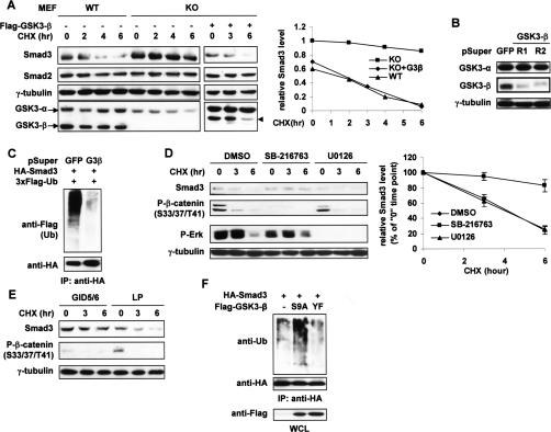
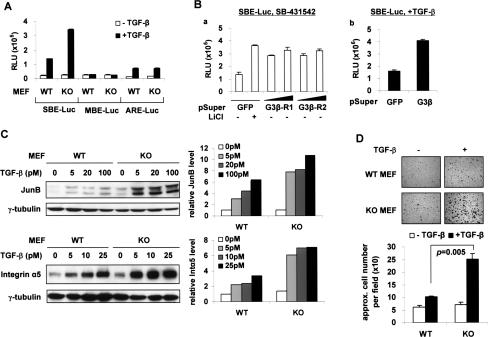
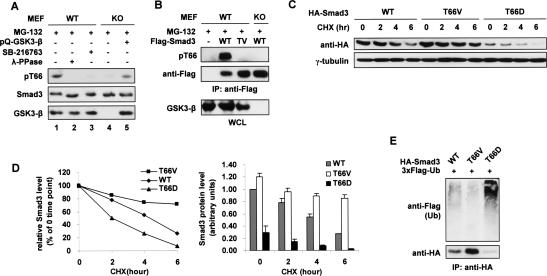
The broad range of biological responses elicited by transforming growth factor-beta (TGF-beta) in various types of tissues and cells is mainly determined by the expression level and activity of the effector proteins Smad2 and Smad3. It is not fully understood how the baseline properties of Smad3 are regulated, although this molecule is in complex with many other proteins at the steady state. Here we show that nonactivated Smad3, but not Smad2, undergoes proteasome-dependent degradation due to the concerted action of the scaffolding protein Axin and its associated kinase, glycogen synthase kinase 3-beta (GSK3-beta). Smad3 physically interacts with Axin and GSK3-beta only in the absence of TGF-beta. Reduction in the expression or activity of Axin/GSK3-beta leads to increased Smad3 stability and transcriptional activity without affecting TGF-beta receptors or Smad2, whereas overexpression of these proteins promotes Smad3 basal degradation and desensitizes cells to TGF-beta. Mechanistically, Axin facilitates GSK3-beta-mediated phosphorylation of Smad3 at Thr66, which triggers Smad3 ubiquitination and degradation. Thr66 mutants of Smad3 show altered protein stability and hence transcriptional activity. These results indicate that the steady-state stability of Smad3 is an important determinant of cellular sensitivity to TGF-beta, and suggest a new function of the Axin/GSK3-beta complex in modulating critical TGF-beta/Smad3-regulated processes during development and tumor progression.


Figures

Figure 1.
Figure 1.
Constitutive proteasomal degradation of steady-state Smad3 is regulated by Axin. (A) HaCaT cells were pretreated with 10 μM SB-431542 for 6 h, and 50 μg/mL CHX or 25 μM MG-132 was then added as indicated. Total cell lysates were probed for endogenous Smad3 and Smad2. γ-Tubulin was used as a loading control. (B) A ubiquitination assay of endogenous Smad3. HaCaT cells were pretreated with either vehicle (ethanol) or 10 μM SB-431542 for 1 h before the addition of 30 μM MG-132. After another 3 h, cells were lysed in SDS lysis buffer for immunoprecipitation (IP) with no antibody (−) or anti-Smad3 (Zymed). (C) Retrovirally infected stable populations of SNU475 cells expressing vector control or wild-type hAxin were pretreated with 10 μM SB-431542 and then treated with 50 μg/mL CHX for the indicated lengths of time. Endogenous proteins were probed. (D) Quantification of endogenous Smad3 level in C and in Alexander and DLD-1 cells tested in similar experiments with the presence of SB-431542. The level of Smad3 in each cell line at time “0” was set as 1.0. (E) Effective knockdown of endogenous Axin in 293T cells by two shRNAs designated as R1 and R2. pSuper-GFP was used as a nontargeting shRNA control. (F,G) Smad3 ubiquitination assays in SNU475 stable lines (F) and in 293T cells (G). pSuper-GFP was used as a control for Axin RNAi (R1 + R2). Cells were pretreated with 10 μM SB-431542 for 1 h followed by MG-132 treatment (25 μM) for 4 h and then lysed in RIPA buffer. Note in G that endogenous hAxin migrates faster than myc-mAxin due to smaller size.
Figure 2.
Figure 2.
Axin negatively affects Smad3-mediated TGF-β activity. (A) Luciferase reporter assays in HepG2 cells. The indicated luciferase constructs and pCMV-β-galactosidase (0.5 μg each) were cotransfected with a total of 3 μg of the pSuper plasmids into each well of a six-well plate, except that, in panel a, 3 and 6 μg of pSuper-Ax-R1 or pSuper-Ax-R2 were used. In panels b and c, mixtures of the two corresponding pSuper-R1 and pSuper-R2 constructs were used, 1.5 μg each. Twenty-four hours post-transfection, cells in panels b and c were incubated with or without 100 pM TGF-β for another 16–24 h. (B) HaCaT cells were infected twice with pSuperRetro viruses. pSR-GL2 targets luciferase and was used as a negative control. Cells were treated with 50 pM TGF-β for 4 h. The protein level of each Smad3 target gene was quantified on the right. (C) HaCaT cells prepared as in B were tested in a wound-healing assay. Cell migration was quantified on the right. (*) P < 0.01. (D) A cell proliferation assay of the indicated HaCaT cells treated with 0, 2.5, 10, 25, or 50 pM TGF-β for 12 h. The P-values between the two types of cells under each concentration of TGF-β are shown. (E) Axin knockdown in HCT116 + Chr.3 and TGF-β treatment were performed as in B. (F) A wound-healing assay of HCT116 + Chr.3 cells. Cells were scratched at 80% confluence, treated with 200 pM TGF-β, and allowed to migrate for 24 h.
Figure 3.
Figure 3.
GSK3-β interacts with nonactivated Smad3. (A) HaCaT cells were pretreated with 50 μM MG-132 for 4 h and with or without 100 pM TGF-β for the last 2 h. Cells were then lysed in ULB+ for endogenous coimmunoprecipitation assays using either preimmune goat IgG or the indicated polyclonal antibodies. Smad2 and Smad3 were probed together with anti-Smad123(H-2). In lanes 4 and 7, ULB+ alone (no lysate) was used as a negative control. (B) Endogenous Smad3 was precipitated from wild-type (WT) and GSK3-β KO MEFs using an anti-Smad3 antibody (Zymed). Coprecipitated GSK3 isoforms were probed. (C) SNU475-Vec or SNU475-Axin cell lysates (1 mg per reaction) were first mixed with 3 μg of GST alone or 1, 3, or 10 μg of GST-Smad3 for 2 h at 4°C and then subjected to anti-GSK3-β immunoprecipitation. GSK3-β did not bind the GST moiety.
Figure 4.
Figure 4.
GSK3-β kinase activity is required for Smad3 basal degradation. (A) Wild-type (WT) and GSK3-β-null MEFs were treated with 50 μg/mL CHX for the indicated time course. Transfected Flag-GSK3-β(WT) is indicated (arrowhead). Protein levels of endogenous Smad3 were analyzed by Western blot and quantified on the right. Smad3 level in the KO cells at time “0” was defined as 1.0. (B) Specific knockdown of GSK3-β in 293T cells by two shRNAs designated as R1 and R2. (C) 293T cells were pretransfected twice with a mixture of the two shRNAs against GSK3-β. After treatment with 10 μM SB-431542 and 25 μM MG-132 for 3 h, cells were lysed in RIPA buffer for the Smad3 ubiquitination assay. (D) HaCaT cells were treated with DMSO, SB-216763 (10 μM), or U0126 (10 μM) for a total of 12 h and with 20 μg/mL CHX for the time course shown. The turnover of endogenous Smad3 was determined by Western blot (left, representative of three independent experiments) and quantified for each treatment group (right). Phospho-β-catenin and phospho-Erk were also examined to show the efficacy of the kinase inhibitors. (E) 293T cells were transfected twice with myc-GID5/6 or the mutant control (myc-GID5/6 LP) before CHX treatment. Endogenous Smad3 and phospho-β-catenin were probed. (F) HA-Smad3 was overexpressed in 293T cells with vector or GSK3-β mutants (S9A or Y216F). After MG-132 treatment (30 μM, 3 h), cells were harvested in RIPA buffer for the Smad3 ubiquitination assay.
Figure 5.
Figure 5.
Selective potentiation of Smad3 activity in GSK3−/− MEFs. (A) MEFs were transfected with the indicated reporters (0.3 μg of SBE/MBE or 0.5 μg of ARE + FoxH1 per well of six-well plates) for 24 h followed by 100 pM TGF-β treatment for 12 h. (B) HepG2 cells were transfected as in Figure 2A. Three micrograms and 6 μg of pSuper-GSK3-β-R1 or pSuper-GSK3-β-R2 were used in panel a, and a mixture of both shRNAs (1.5 μg each) was used in panel b. LiCl (20 mM) and TGF-β (100 pM) treatments both lasted for 24 h. (C) Wild-type (WT) and KO MEFs were treated with different concentrations of TGF-β for 2 h and harvested in ULB+. Protein expressions were analyzed by Western blot (left) and quantified (right). (D) A cell adhesion assay of the MEFs. The numbers of attached cells were quantified and presented as mean ± SD.
Figure 6.
Figure 6.
GSK3-β-mediated Thr66 phosphorylation leads to Smad3 basal degradation. (A) Endogenous Smad3 is phosphorylated at Thr66. GSK3-β wild-type (WT) and KO MEFs were treated with 20 μM MG-132 for 3 h, and cell lysates were probed with the anti-pT66 antiserum. (Lane 3) SB-216763 (10 μM) was added 10 h prior to MG-132 treatment. (Lane 5) pQCXIP-GSK3-β(WT) was transfected 24 h prior to cell lysis. (B) GSK3-β wild-type (WT) and KO MEFs were transfected with 3xFlag-Smad3(WT or T66V) and treated with MG-132 as in A. Cells were harvested in RIPA buffer followed by anti-Flag IP. (C) Equal amounts of HA-tagged Smad3 constructs (WT, T66V, or T66D) were individually expressed in 293T cells for 20 h before the indicated CHX treatment (50 μg/mL). The Western blot is representative of three independent experiments, and the results from these experiments are quantitatively presented in D to show the turnover rate (left) and the protein level (right) of the Smad3 variants. (E) 293T cells transfected with the indicated constructs were treated with 30 μM MG-132 for 3 h and lysed in SDS lysis buffer for the ubiquitination assay.
Figure 7.
Figure 7.
Smad3(T66V) is hyperactive. (A) HepG2 cells seeded in 12-well plates were transfected with the indicated amounts of 3xFlag-Smad3(WT or T66V). Cells were treated with or without TGF-β (200 pM) for 12 h and assessed for proliferation. The data are presented as the percentage of growth inhibition compared with vector-transfected, untreated cells. (B) An equal amount of vector, HA-Smad3(WT), or HA-Smad3(T66V) was transfected into GSK3-β wild-type (WT) and KO MEFs. Twenty-four hours later, cells were treated with 25 pM TGF-β for 4 h. (Right) The protein levels of Integrin α5 and JunB were quantified. (C) SMAD3−/− MEFs were transfected with 1 μg of vector control or 3xFlag-Smad3(WT, T66V, or T66D). Twelve hours post-transfection, 10 μM SB-216763 or DMSO was added. Cells were harvested in ULB+ after another 12 h. (D) An SBE-Luc reporter assay in SMAD3−/− MEFs. After transfection of the luciferase construct, cells were incubated with or without 20 mM LiCl for 24 h. (E) A schematic representation of the Smad3/Axin/GSK3-β complex (right) in comparison with key components of the β-catenin destruction complex (left). Unlike Wnt, TGF-β does not disassemble the Axin/GSK3-β complex. The rapid nuclear translocation of Smad3 is caused by the C-terminal phosphorylation rather than protein accumulation.

References

    1. Bierie B., Moses H.L. Tumour microenvironment: TGFβ: The molecular Jekyll and Hyde of cancer. Nat. Rev. Cancer. 2006;6:506–520. - PubMed
    1. Chen H.B., Shen J., Ip Y.T., Xu L. Identification of phosphatases for Smad in the BMP/DPP pathway. Genes & Dev. 2006;20:648–653. - PMC - PubMed
    1. Clarke D.C., Betterton M.D., Liu X. Systems theory of Smad signalling. IEE Proc. Syst. Biol. 2006;153:412–424. - PubMed
    1. Cohen P., Frame S. The renaissance of GSK3. Nat. Rev. Mol. Cell Biol. 2001;2:769–776. - PubMed
    1. Cross D.A., Alessi D.R., Cohen P., Andjelkovich M., Hemmings B.A. Inhibition of glycogen synthase kinase-3 by insulin mediated by protein kinase B. Nature. 1995;378:785–789. - PubMed

Publication types

MeSH terms

LinkOut - more resources