Ca2+ channel-independent requirement for MAGUK family CACNB4 genes in initiation of zebrafish epiboly
- PMID: 18172207
- PMCID: PMC2224185
- DOI: 10.1073/pnas.0707948105
Ca2+ channel-independent requirement for MAGUK family CACNB4 genes in initiation of zebrafish epiboly
Abstract
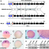
CACNB genes encode membrane-associated guanylate kinase (MAGUK) proteins once thought to function exclusively as auxiliary beta subunits in assembly and gating of voltage-gated Ca(2+) channels. Here, we report that zygotic deficiency of zebrafish beta4 protein blocks initiation of epiboly, the first morphogenetic movement of teleost embryos. Reduced beta4 function in the yolk syncytial layer (YSL) leads to abnormal division and dispersal of yolk syncytial nuclei, blastoderm retraction, and death, effects highly similar to microtubule disruption by nocodazole. Epiboly is restored by coinjection of human beta4 cRNA or, surprisingly, by mutant cRNA encoding beta4 subunits incapable of binding to Ca(2+) channel alpha1 subunits. This study defines a YSL-driven zygotic mechanism essential for epiboly initiation and reveals a Ca(2+) channel-independent beta4 protein function potentially involving the cytoskeleton.
Conflict of interest statement
The authors declare no conflict of interest.
Figures


Similar articles
-
Genomic organization, expression, and phylogenetic analysis of Ca2+ channel beta4 genes in 13 vertebrate species.Physiol Genomics. 2008 Oct 8;35(2):133-44. doi: 10.1152/physiolgenomics.90264.2008. Epub 2008 Aug 5. Physiol Genomics. 2008. PMID: 18682574 Free PMC article.
-
Microtubule arrays of the zebrafish yolk cell: organization and function during epiboly.Development. 1994 Sep;120(9):2443-55. doi: 10.1242/dev.120.9.2443. Development. 1994. PMID: 7956824
-
The calcium channel beta2 (CACNB2) subunit repertoire in teleosts.BMC Mol Biol. 2008 Apr 17;9:38. doi: 10.1186/1471-2199-9-38. BMC Mol Biol. 2008. PMID: 18419826 Free PMC article.
-
Zebrafish epiboly: Spreading thin over the yolk.Dev Dyn. 2016 Mar;245(3):244-58. doi: 10.1002/dvdy.24353. Epub 2015 Oct 28. Dev Dyn. 2016. PMID: 26434660 Review.
-
Functional diversity in neuronal voltage-gated calcium channels by alternative splicing of Ca(v)alpha1.Mol Neurobiol. 2002 Aug;26(1):21-44. doi: 10.1385/MN:26:1:021. Mol Neurobiol. 2002. PMID: 12392054 Review.
Cited by
-
Cloning and characterization of voltage-gated calcium channel alpha1 subunits in Xenopus laevis during development.Dev Dyn. 2009 Nov;238(11):2891-902. doi: 10.1002/dvdy.22102. Dev Dyn. 2009. PMID: 19795515 Free PMC article.
-
Adult and Developing Zebrafish as Suitable Models for Cardiac Electrophysiology and Pathology in Research and Industry.Front Physiol. 2021 Jan 13;11:607860. doi: 10.3389/fphys.2020.607860. eCollection 2020. Front Physiol. 2021. PMID: 33519514 Free PMC article. Review.
-
Zebrafish Models of Autosomal Dominant Ataxias.Cells. 2021 Feb 17;10(2):421. doi: 10.3390/cells10020421. Cells. 2021. PMID: 33671313 Free PMC article. Review.
-
The ß subunit of voltage-gated Ca2+ channels.Physiol Rev. 2010 Oct;90(4):1461-506. doi: 10.1152/physrev.00057.2009. Physiol Rev. 2010. PMID: 20959621 Free PMC article. Review.
-
ApoA-II directs morphogenetic movements of zebrafish embryo by preventing chromosome fusion during nuclear division in yolk syncytial layer.J Biol Chem. 2011 Mar 18;286(11):9514-25. doi: 10.1074/jbc.M110.134908. Epub 2011 Jan 6. J Biol Chem. 2011. PMID: 21212265 Free PMC article.
References
-
- Solnica-Krezel L. Curr Opin Genet Dev. 2006;16:433–441. - PubMed
-
- Rohde LA, Heisenberg CP. Int Rev Cytol. 2007;261C:159–192. - PubMed
-
- Cheng JC, Miller AL, Webb SE. Dev Dyn. 2004;231:313–323. - PubMed
-
- Zalik SE, Lewandowski E, Kam Z, Geiger B. Biochem Cell Biol. 1999;77:527–542. - PubMed
-
- Solnica-Krezel L, Driever W. Development. 1994;120:2443–2455. - PubMed
Publication types
MeSH terms
Substances
Associated data
- Actions
- Actions
- Actions
- Actions
- Actions
- Actions
Grants and funding
LinkOut - more resources
Full Text Sources
Other Literature Sources
Molecular Biology Databases
Miscellaneous

