. 2008 Jan 8;105(1):186-91.
doi: 10.1073/pnas.0706183104. Epub 2008 Jan 2.

Dioxin-mediated tumor progression through activation of mitochondria-to-nucleus stress signaling

Affiliations

Dioxin-mediated tumor progression through activation of mitochondria-to-nucleus stress signaling

Gopa Biswas et al. Proc Natl Acad Sci U S A. .

Abstract

The environmental toxin 2,3,7,8-tetrachlorodibenzodioxin (TCDD) is a known human carcinogen; however, its precise mechanism of action remains unclear. Here we show that TCDD induces mitochondrial dysfunction, stress signaling, and tumor invasion by a mechanism similar to that described for mtDNA-depleted cells. Treatment of C2C12 cells with TCDD disrupted mitochondrial transmembrane potential in a time-dependent fashion and inhibited mitochondrial transcription and translation. TCDD also increased cytosolic [Ca(2+)](c) and RyR1-specific Ca(2+) release. These changes were associated with increased calcineurin (CnA) levels and activation of CnA-sensitive NF-kappaB/Rel (IkappaBbeta-dependent) factors. Cells treated with TCDD displayed resistance to apoptosis, increased expression of the tumor marker cathepsin L, and a high degree of invasiveness as tested by the Matrigel membrane invasion assay. These effects were reversed by the CnA inhibitor FK506, and CnA mRNA silencing suggesting that TCDD triggers a signaling pathway similar to mtDNA depletion. Taken together, these results reveal that TCDD may promote tumor progression in vivo by directly targeting mitochondrial transcription and induction of mitochondrial stress signaling.


Conflict of interest statement

The authors declare no conflict of interest.

Figures
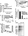
Fig. 1.
Fig. 1.
TCDD disrupts ΔΨm and alters Ca2+ homeostasis in C2C12 cells. (A) Uptake of MitoTracker Orange was recorded in control (●), mtDNA-depleted (■), CCCP-treated (▴), or TCDD-treated (10 nM) (×) cultures. (B and C) Ca2+ release was measured with 1 μM Fura 2FF/FA by exposing control and TCDD-treated cells to 10 μM acetylcholine (B) and 20 mM caffeine (C), respectively. Where indicated, cultures were treated with 100 mM KCl to restore ΔΨm. (D) Mitochondrial genome-coded CcOI and CcOII and nuclear genome-coded CcOIVi1 mRNA levels were quantified by real-time PCR with β-actin gene as internal control. Treatment with 10 nM TCDD was for 4 h. (E) Level of mitochondrial transcription in isolated mitochondria. [32P]UTP incorporation was carried out as described in Methods. (F) Mitochondrial translation products were labeled with [35S]Met as described in Methods. Proteins were analyzed by SDS/PAGE followed by fluorography.
Fig. 2.
Fig. 2.
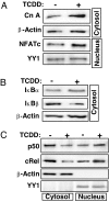
TCDD induces expression of mitochondrial stress signaling marker proteins. Subcellular fractions isolated from control and 10 nM TCDD-treated cells were analyzed by Western blotting for CnA/NFATc (A), IκBα and IκBβ (B), and NF-κB transcription factors (C). β-Actin and YY1 were used as loading controls for cytosolic and nuclear fractions, respectively.
Fig. 3.
Fig. 3.
TCDD increases cathepsin L and RyR1 levels, promotes invasive behavior, and confers resistance to apoptosis in a CnA-dependent manner. (A) Western blot analysis of cathepsin L and RyR1 levels in whole-cell lysates from control, CnA-silenced (siCnA), TCDD-treated, and TCDD + FK506-treated cells. Na+/K+-ATPase served as a loading control. (B) A Matrigel invasion assay was performed on control, CnAα-silenced (siCnAα), TCDD-treated, and TCDD + FK506-treated cells. Invaded cells were stained and viewed as described in Methods. Numbers in parentheses underneath the images indicate the number of cells in the area shown. (C) The effect of TCDD on STP-induced apoptotic cell death was assessed by TUNEL assay.
Fig. 4.
Fig. 4.
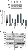
TCDD-induced mitochondrial stress signaling is unaffected by AhR inhibition and Arnt mRNA silencing. (A) Nuclear fractions isolated from control (Cont), mtDNA-depleted (Depl), and TCDD-treated cells were subjected to Western blot analysis of AhR and Arnt. MCF7 nuclear fractions were included as a positive control. YY1 was used as a loading control. (B) Arnt mRNA levels were assessed by real-time PCR in control cells, TCDD-treated cells, mtDNA-depleted cells, Arnt-silenced control cells, and Arnt-silenced mtDNA-depleted cells. (C) Effect of α-NF, an AhR antagonist, and Arnt mRNA silencing on the induction of mitochondrial stress signaling markers by TCDD. Levels of the indicated factors were assessed by Western blot analysis. YY1 and β-actin were used as loading controls for nuclear and cytosolic fractions, respectively.
Fig. 5.
Fig. 5.
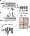
TCDD-induced expression of marker genes in Arnt mRNA-silenced cells. (A) Cathepsin L and RyR1 protein levels in postnuclear fractions were analyzed by Western blotting. (B) immunoblot of cathepsin L and RyR1 levels in Arnt+/+ and Arnt−/− MEFs as in A. (C) Levels of RyR1 mRNA (Upper) and cathepsin L mRNA (CathL; Lower) were assessed by RT-PCR amplification followed by Southern blot hybridization and real-time PCR, respectively. β-Actin served as an internal control. (D) Control cells, TCDD-treated cells, mtDNA-depleted cells, Arnt silenced control cells, and Arnt-silenced mtDNA-depleted cells were evaluated for invasiveness in response to TCDD treatment by using a Matrigel invasion assay.

Similar articles

Cited by

References

    1. Wallace DC. Annu Rev Genet. 2005;39:359–407. - PMC - PubMed
    1. Singh KK, Russell J, Sigala B, Zhang Y, Williams J, Keshav KF. Oncogene. 1999;18:6641–6646. - PubMed
    1. Amuthan G, Biswas G, Zhang SY, Klein-Szanto A, Vijayasarathy C, Avadhani NG. EMBO J. 2001;20:1910–1920. - PMC - PubMed
    1. Bandy B, Davison AJ. Free Radic Biol Med. 1990;8:523–539. - PubMed
    1. Niranjan BG, Bhat NK, Avadhani NG. Science. 1982;215:73–75. - PubMed

Publication types

LinkOut - more resources