. 2008 Jan 1;68(1):89-97.
doi: 10.1158/0008-5472.CAN-07-0675.

DNA protein kinase-dependent G2 checkpoint revealed following knockdown of ataxia-telangiectasia mutated in human mammary epithelial cells

Affiliations

DNA protein kinase-dependent G2 checkpoint revealed following knockdown of ataxia-telangiectasia mutated in human mammary epithelial cells

Sonnet J H Arlander et al. Cancer Res. .

Abstract

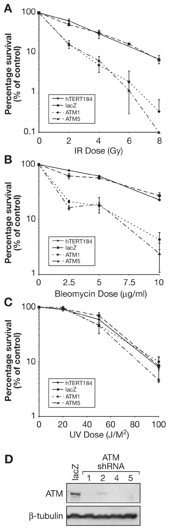
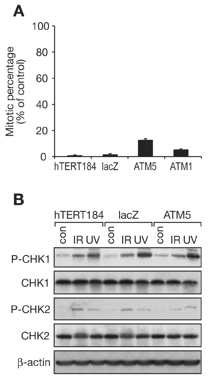
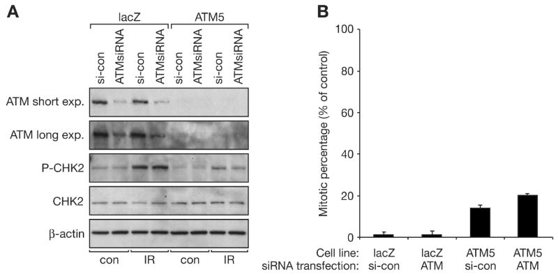
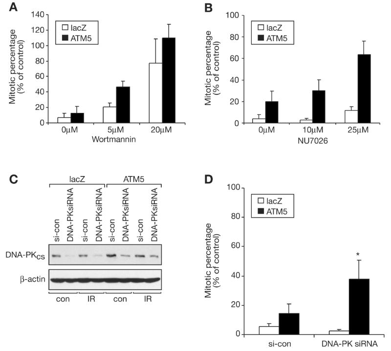
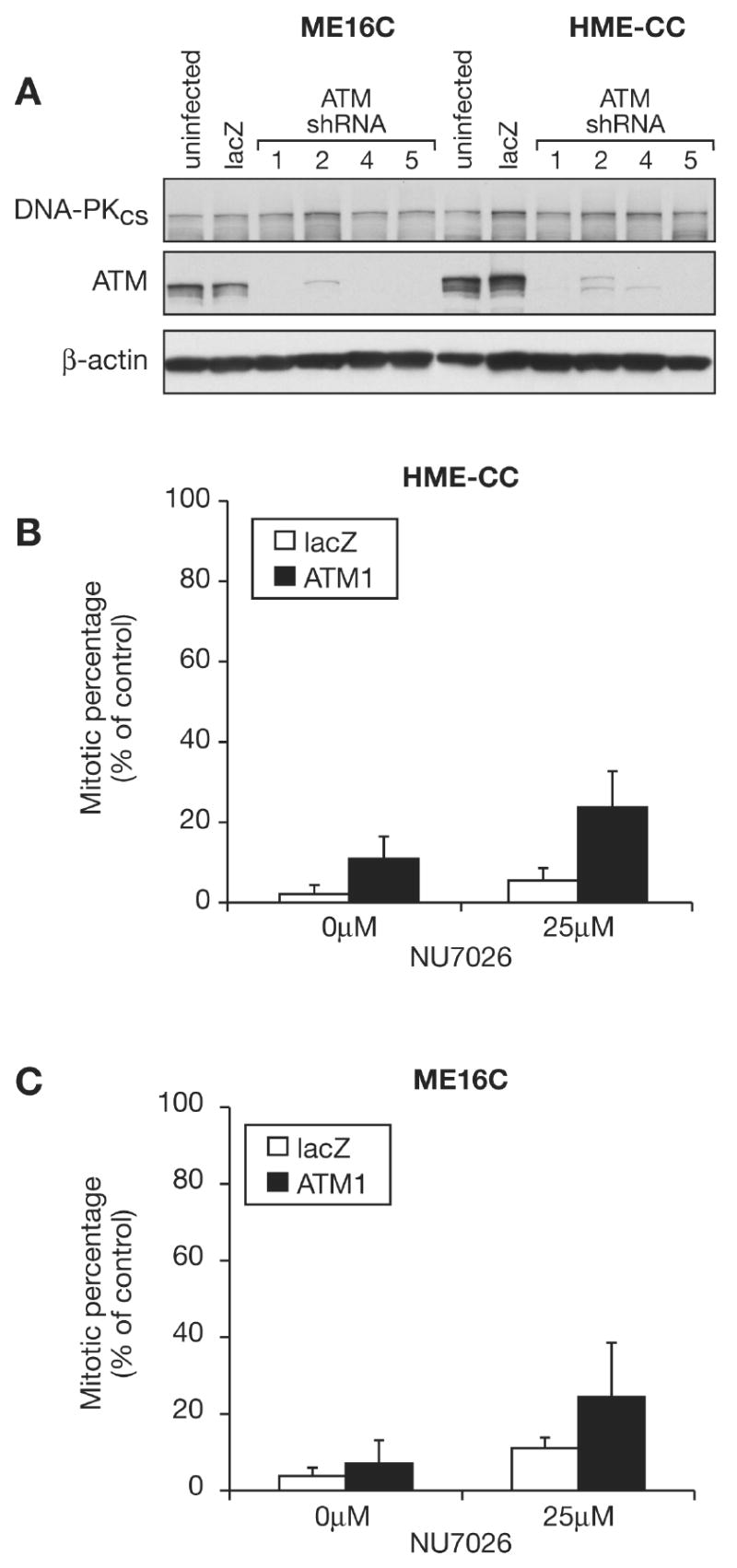
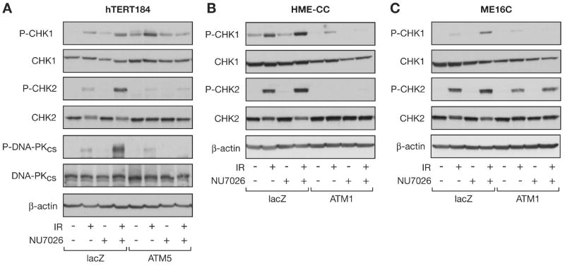
Members of the phosphatidylinositol 3-kinase-related kinase family, in particular the ataxia-telangiectasia mutated (ATM) kinase and the catalytic subunit of the DNA-dependent protein kinase (DNA-PKcs), regulate cellular responses to DNA double-strand breaks. Increased sensitivity to ionizing radiation (IR) in DNA-PKcs- or ATM-deficient cells emphasizes their important roles in maintaining genome stability. Furthermore, combined knockout of both kinases is synthetically lethal, suggesting functional complementarity. In the current study, using human mammary epithelial cells with ATM levels stably knocked down by >90%, we observed an IR-induced G(2) checkpoint that was only slightly attenuated. In marked contrast, this G(2) checkpoint was significantly attenuated with either DNA-PK inhibitor treatment or RNA interference knockdown of DNA-PKcs, the catalytic subunit of DNA-PK, indicating that DNA-PK contributes to the G(2) checkpoint in these cells. Furthermore, in agreement with the checkpoint attenuation, DNA-PK inhibition in ATM-knockdown cells resulted in reduced signaling of the checkpoint kinase CHK1 as evidenced by reduced CHK1 phosphorylation. Taken together, these results show a DNA-PK-dependent component to the IR-induced G(2) checkpoint, in addition to the well-defined ATM-dependent component. This may have important implications for chemotherapeutic strategies for breast cancers.


Figures

Figure 1
Figure 1
Clonogenic survival shows that stable knockdown of ATM in hTERT184 cells sensitizes cells to IR and bleomyocin sulfate but not UV radiation treatment. hTERT184 cells and derivative lines were treated with the indicated amounts of A. IR B. bleomycin sulfate or C. UV radiation and colonies allowed to grow for 10 days. Results are the mean ± SEM of at least 3 independent experiments in duplicate plates. D. Human mammary epithelial hTERT184 cells were infected with lentiviruses encoding shRNAs against ATM (#1, 2, 4, and 5) and stable transfectants selected in 4 μg/ml blasticidin. Western blot analysis of ATM levels was performed with β-tubulin used as a loading control.
Figure 2
Figure 2. ATM knockdown hTERT184 cells demonstrate a G2 checkpoint response following IR and IR-induced phosphorylation of CHK1 and CHK2 in ATM knockdown cells
A. hTERT184-lacZ, -ATM5, and -ATM1 cells were treated with 2 Gy IR and collected and fixed 2 h after IR treatment. Cells were stained with a phospho-serine10 histone H3 antibody to assess the G2 checkpoint by flow cytometry. Mean mitotic percentages ± s.d. of 3 independent experiments. B. Western blot analysis of phosphorylation of CHK1 and CHK2 after IR in hTERT184 and derivative cells. Cells were treated with 2 Gy IR, cultured for an additional 2 h and then analyzed for phospho-CHK1 (Ser345), phospho-CHK2 (Thr68), total CHK1 and total CHK2 levels. β-actin was used as a loading control. As a positive control for CHK1 phosphorylation, cells were also treated with 50 J/M2 UV radiation followed by culturing for an additional 2 h. (con = control)
Figure 3
Figure 3. Transient transfection of hTERT184-ATM5 stable knockdown cell line with ATM siRNA only slightly attenuates the G2 checkpoint response
A. hTERT184-lacZ and hTERT184-ATM5 cells were transfected with a control siRNA or a siRNA against ATM for 48 h, exposed to 2 Gy IR and then cultured for an additional 2 h. Western blot analysis of ATM, phospho-CHK2 (Thr68), and total CHK2 levels was performed. β-actin was used as a loading control. B. Cells treated as in A., but 2 h after IR, cells were collected and fixed. Flow cytometry was performed using a phospho-histone H3 (Ser10) antibody to assess the G2 checkpoint. Data shown are the mean mitotic percentages ± s.d. of 2 independent experiments. (con = control, si-con = control siRNA)
Figure 4
Figure 4. Wortmannin, the DNA-PK inhibitor NU7026, and DNA-PKcs siRNA attenuate the IR-induced G2 checkpoint response in hTERT184 cells
A, B. Cells were pretreated with the indicated amounts of wortmannin or NU7026 1 h prior to 2 Gy IR treatment. Cells were collected and fixed 2 h after IR. Flow cytometry was performed using a phospho-histone H3 (Ser10) antibody to assess the IR-induced G2 checkpoint. Data shown are the mean mitotic percentages ± s.d. of 2 or 3 independent experiments. C. hTERT184-lacZ and hTERT184-ATM5 cells were transfected with a control siRNA (si-con) or a siRNA against DNA-PKcs (DNA-PKsiRNA) for 48 h, then treated with 2 Gy IR and cultured for an additional 2 h. Western blot analysis of DNA-PK with β-actin a loading control. D. Cells were treated as in C., but 2 h after IR, cells were collected and fixed. Flow cytometry was performed using a phospho-serine 10 histone H3 antibody to assess the IR-induced G2 checkpoint. Data shown are the mean mitotic percentages ± s.d. of 3 independent experiments. *, p < 0.05 relative to transfection with control siRNA.
Figure 5
Figure 5. DNA-PK inhibition with NU7026 attenuates the IR-induced G2 checkpoint response in HME-CC and ME16C human mammary epithelial cells
A. Human mammary epithelial ME16C and HME-CC cells were infected with lentiviruses encoding shRNAs against ATM (#1, 2, 4, and 5) and stable transfectants selected in 4 μg/ml blasticidin. Western blot analysis of ATM and DNA-PKcs levels were performed with β-actin used as a loading control. B, C. HME-CC and ME16C cells, respectively, were pretreated with the indicated amounts of NU7026 1 h prior to 2 Gy IR treatment. Cells were collected and fixed 2 h after IR. Flow cytometry was performed using a phospho-histone H3 (Ser10) antibody to assess the IR-induced G2 checkpoint. Data shown are the mean mitotic percentages ± s.d. of 3 independent experiments.
Figure 6
Figure 6
IR-induced CHK1 phosphorylation is inhibited by the DNA-PK inhibitor NU7026 in hTERT184, HME-CC, and ME16C cells with reduced ATM levels. Western blot analysis of phosphorylation of CHK1 and CHK2 after IR in A. hTERT184, B. HME-CC, and C. ME16C cells. Cells were pretreated with 25 μM of NU7026, as indicated, 1 h prior to 2 Gy IR treatment, cultured for an additional 2 h and then analyzed for phospho-CHK1, phospho-CHK2, total CHK1 and total CHK2 levels. Total DNA-PKcs and phospho-DNA-PK levels are shown in hTERT184 lines. β-actin was used as a loading control.

Similar articles

Cited by

References

    1. Niida H, Nakanishi M. DNA damage checkpoints in mammals. Mutagenesis. 2006;21(1):3–9. - PubMed
    1. Meek K, Gupta S, Ramsden DA, Lees-Miller SP. The DNA-dependent protein kinase: the director at the end. Immunol Rev. 2004;200:132–41. - PubMed
    1. Lavin MF, Birrell G, Chen P, Kozlov S, Scott S, Gueven N. ATM signaling and genomic stability in response to DNA damage. Mutat Res. 2005;569(12):123–32. - PubMed
    1. Brown EJ, Baltimore D. ATR disruption leads to chromosomal fragmentation and early embryonic lethality. Genes Dev. 2000;14(4):397–402. - PMC - PubMed
    1. Shinohara ET, Geng L, Tan J, et al. DNA-dependent protein kinase is a molecular target for the development of noncytotoxic radiation-sensitizing drugs. Cancer Res. 2005;65(12):4987–92. - PubMed

Publication types

MeSH terms

Substances