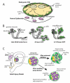
The scoop on the fly brain: glial engulfment functions in Drosophila
- PMID: 18172512
- PMCID: PMC2171361
- DOI: 10.1017/S1740925X07000646
The scoop on the fly brain: glial engulfment functions in Drosophila
Abstract
Glial cells provide support and protection for neurons in the embryonic and adult brain, mediated in part through the phagocytic activity of glia. Glial cells engulf apoptotic cells and pruned neurites from the developing nervous system, and also clear degenerating neuronal debris from the adult brain after neural trauma. Studies indicate that Drosophila melanogaster is an ideal model system to elucidate the mechanisms of engulfment by glia. The recent studies reviewed here show that many features of glial engulfment are conserved across species and argue that work in Drosophila will provide valuable cellular and molecular insight into glial engulfment activity in mammals.
Keywords: Drosophila glia; Phagocyte; phagocytosis.
Figures


Similar articles
-
Neuron-glia crosstalk in neuronal remodeling and degeneration: Neuronal signals inducing glial cell phagocytic transformation in Drosophila.Bioessays. 2022 May;44(5):e2100254. doi: 10.1002/bies.202100254. Epub 2022 Mar 22. Bioessays. 2022. PMID: 35315125
-
Glia actively sculpt sensory neurons by controlled phagocytosis to tune animal behavior.Elife. 2021 Mar 24;10:e63532. doi: 10.7554/eLife.63532. Elife. 2021. PMID: 33759761 Free PMC article.
-
Astrocytes engage unique molecular programs to engulf pruned neuronal debris from distinct subsets of neurons.Genes Dev. 2014 Jan 1;28(1):20-33. doi: 10.1101/gad.229518.113. Epub 2013 Dec 20. Genes Dev. 2014. PMID: 24361692 Free PMC article.
-
Keeping the CNS clear: glial phagocytic functions in Drosophila.Glia. 2011 Sep;59(9):1304-11. doi: 10.1002/glia.21098. Epub 2010 Dec 6. Glia. 2011. PMID: 21136555 Review.
-
Engulfed by Glia: Glial Pruning in Development, Function, and Injury across Species.J Neurosci. 2021 Feb 3;41(5):823-833. doi: 10.1523/JNEUROSCI.1660-20.2020. Epub 2021 Jan 19. J Neurosci. 2021. PMID: 33468571 Free PMC article. Review.
Cited by
-
Myelination transition zone astrocytes are constitutively phagocytic and have synuclein dependent reactivity in glaucoma.Proc Natl Acad Sci U S A. 2011 Jan 18;108(3):1176-81. doi: 10.1073/pnas.1013965108. Epub 2011 Jan 3. Proc Natl Acad Sci U S A. 2011. PMID: 21199938 Free PMC article.
-
Negative regulation of glial engulfment activity by Draper terminates glial responses to axon injury.Nat Neurosci. 2012 Mar 18;15(5):722-30. doi: 10.1038/nn.3066. Nat Neurosci. 2012. PMID: 22426252 Free PMC article.
-
Delayed glial clearance of degenerating axons in aged Drosophila is due to reduced PI3K/Draper activity.Nat Commun. 2016 Sep 20;7:12871. doi: 10.1038/ncomms12871. Nat Commun. 2016. PMID: 27647497 Free PMC article.
-
Mechanisms governing activity-dependent synaptic pruning in the developing mammalian CNS.Nat Rev Neurosci. 2021 Nov;22(11):657-673. doi: 10.1038/s41583-021-00507-y. Epub 2021 Sep 20. Nat Rev Neurosci. 2021. PMID: 34545240 Free PMC article. Review.
-
Protein phosphatase 4 coordinates glial membrane recruitment and phagocytic clearance of degenerating axons in Drosophila.Cell Death Dis. 2017 Feb 23;8(2):e2623. doi: 10.1038/cddis.2017.40. Cell Death Dis. 2017. PMID: 28230857 Free PMC article.
References
-
- Abrams JM, White K, Fessler LI, Steller H. Programmed cell death during Drosophila embryogenesis. Development. 1993;117:29–43. - PubMed
-
- Allen NJ, Barres BA. Signaling between glia and neurons: focus on synaptic plasticity. Current Opinion in Neurobiology. 2005;15:542–548. - PubMed
-
- Awasaki T, Ito K. Engulfing action of glial cells is required for programmed axon pruning during Drosophila metamorphosis. Current Biology. 2004;14:668–677. - PubMed
-
- Awasaki T, Tatsumi R, Takahashi K, Arai K, Nakanishi Y, Ueda R, Ito K. Essential role of the apoptotic cell engulfment genes draper and ced-6 in programmed axon pruning during Drosophila metamorphosis. Neuron. 2006;50:855–867. - PubMed
-
- Bangs P, White K. Regulation and execution of apoptosis during Drosophila development. Developmental Dynamics. 2000;218:68–79. - PubMed
Grants and funding
LinkOut - more resources
Full Text Sources
Molecular Biology Databases

