. 2008 Feb;118(2):777-88.
doi: 10.1172/JCI32806.

Dopamine-modified alpha-synuclein blocks chaperone-mediated autophagy

Affiliations

Dopamine-modified alpha-synuclein blocks chaperone-mediated autophagy

Marta Martinez-Vicente et al. J Clin Invest. 2008 Feb.

Abstract

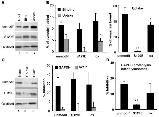
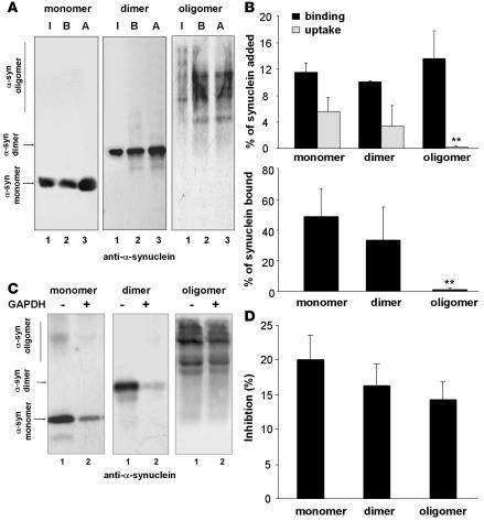
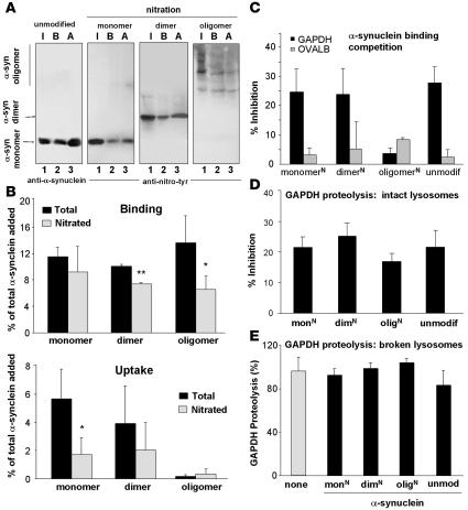
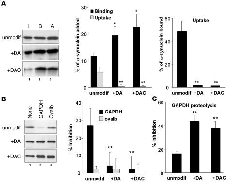
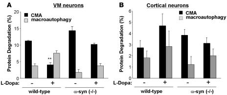
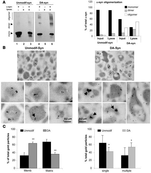
Altered degradation of alpha-synuclein (alpha-syn) has been implicated in the pathogenesis of Parkinson disease (PD). We have shown that alpha-syn can be degraded via chaperone-mediated autophagy (CMA), a selective lysosomal mechanism for degradation of cytosolic proteins. Pathogenic mutants of alpha-syn block lysosomal translocation, impairing their own degradation along with that of other CMA substrates. While pathogenic alpha-syn mutations are rare, alpha-syn undergoes posttranslational modifications, which may underlie its accumulation in cytosolic aggregates in most forms of PD. Using mouse ventral medial neuron cultures, SH-SY5Y cells in culture, and isolated mouse lysosomes, we have found that most of these posttranslational modifications of alpha-syn impair degradation of this protein by CMA but do not affect degradation of other substrates. Dopamine-modified alpha-syn, however, is not only poorly degraded by CMA but also blocks degradation of other substrates by this pathway. As blockage of CMA increases cellular vulnerability to stressors, we propose that dopamine-induced autophagic inhibition could explain the selective degeneration of PD dopaminergic neurons.


Figures

Figure 1
Figure 1. Effect of oxidation and phosphorylation of α-syn on its degradation in lysosomes by CMA.
(A) Association of unmodified and oxidized α-syn and of the S129E mutant of α-syn with isolated lysosomes untreated (binding [Bind]) or previously treated with proteinase inhibitors (association: binding + uptake [Assoc]). Lane 1 shows one-tenth of the amount of protein added to the incubation (Input). (B) Percentage of each protein bound and translocated (uptake = association – binding) inside lysosomes calculated from the densitometric quantification of 11–13 immunoblots such as the representative immunoblots shown in A. The right-hand bars show the percentage of α-syn bound to the lysosomal membrane that was translocated into lysosomes. (C) Effect of a 2-molar excess of GAPDH or ovalbumin (Ovalb) on the association of unmodified, oxidized, and S129E mutant α-syn with lysosomes. Left panel: representative immunoblot. Right panel: percentage of inhibition of the lysosomal association of each form of α-syn calculated from the densitometric quantification of 4–6 immunoblots such as those shown here. (D) Effect of adding these 3 forms of α-syn in equimolar ratio with [14C]GAPDH on the degradation of [14C]GAPDH by intact lysosomes. Values are expressed as percentage of inhibition of GAPDH degradation and are mean + SEM of 4–5 experiments with triplicate samples. *P < 0.05; **P < 0.01.
Figure 2
Figure 2. Degradation in lysosomes by CMA of multimeric forms of α-syn.
(A) Association of monomers and irreversibly cross-linked dimers and oligomers of α-syn with isolated lysosomes untreated (binding [B]) or previously treated with proteinase inhibitors (association [A]). Lane 1 shows one-tenth of the amount of protein added to the incubation (input [I]). (B) Upper panel: percentage of each protein bound and translocated (uptake = association – binding) inside lysosomes, calculated from the densitometric quantification of 6–8 immunoblots as used for the immunoblots shown in A. Lower panel: percentage of α-syn bound to the lysosomal membrane that is translocated into lysosomes. (C) Effect of a 2-molar excess of GAPDH on the association of monomeric, dimeric, or multimeric α-syn with lysosomes. (D) Percentage of inhibition of the lysosomal association of each form of α-syn calculated from the densitometric quantification of 4–6 immunoblots such as those shown in C. **P < 0.01.
Figure 3
Figure 3. Effect of nitration of α-syn on its degradation in lysosomes by CMA.
(A) Association of unmodified α-syn and the monomeric, dimeric, and oligomeric forms resulting from nitration of α-syn with isolated lysosomes untreated (binding [B]) or previously treated with proteinase inhibitors (association: binding + uptake [A]). Input (I): one-fifth of the amount of protein added to the incubation. Blots were developed with an antibody against α-syn (unmodified protein) or one that only recognizes the nitrated forms of α-syn (nitrated samples). (B) Percentage of each protein bound (upper panel) and translocated (lower panel) inside lysosomes, calculated from the densitometric quantification of immunoblots developed with both antibodies. n = 6. (C) Effect of a 2-molar excess of GAPDH on the association of monomers, dimers, and oligomers of nitrated α-syn (N) with lysosomes. Values are expressed as percentage of inhibition of the lysosomal association of each form of α-syn. n = 4–6. (D and E) Effect of equimolar amounts of unmodified monomers (unmodif) and nitrated (N) monomers (mon), dimers (dim), and oligomers (olig) of α-syn on the degradation of [14C]GAPDH by intact (D) or broken (E) lysosomes. Values are expressed as percentage of inhibition of the degradation of GAPDH (D) or as percentage of GAPDH degradation at the end of incubation (E) and are mean + SEM of 4–5 experiments with triplicate samples. *P < 0.05; **P < 0.01.
Figure 4
Figure 4. Degradation of dopamine-reacted α-syn in lysosomes by CMA.
(A) Association of unmodified, dopamine-reacted (+DA) and dopaminochrome-reacted (+DAC) α-syn with isolated lysosomes untreated (binding [B]) or previously treated with proteinase inhibitors (association: binding + uptake [A]). Input (I): one-fifth of the amount of protein added to the incubation. The percentage of each protein bound and translocated inside lysosomes (middle) and the percentage of bound protein translocated into lysosomes (right) was calculated from the densitometric quantification of immunoblots. n = 6–8. (B) Effect of a 2-molar excess of GAPDH or ovalbumin (ovalb) on the association of unmodified and DA– and DAC–α-syn with lysosomes. Values are the percentage of inhibition of the lysosomal association of each form of α-syn. n = 6. (C) Effect of unmodified and DA– and DAC–α-syn in 0.5:1 molar ratio with [14C]GAPDH in the degradation of [14C]GAPDH by intact lysosomes. Values are mean + SEM of the percentage of GAPDH degradation at the end of the incubation in 4–5 experiments with triplicate samples. **P < 0.01.
Figure 5
Figure 5. Dopamine-induced blockage of CMA in VM neuron cultures.
Effect of l-DOPA treatment on the degradation rates of long-lived proteins in cultured dopaminergic neurons (VM neurons) (A) and cortical neurons (B) from wild-type and homozygous (–/–) α-syn–knockout mice. Protein degradation was calculated as the percentage of total protein (acid precipitable radioactivity) at time 0, converted into amino acids and small peptides (acid soluble radioactivity) after 20 hours. The contribution of different autophagies to total protein degradation was calculated as protein degradation sensitive to ammonium chloride and 3-methyladenine (macroautophagy) and protein degradation sensitive to ammonium chloride but insensitive to 3-methyladenine (CMA). n = 3 experiments with at least 6 dishes for each condition. **P < 0.01.
Figure 6
Figure 6. Increased intracellular levels of dopamine enhance the association of modified α-syn to lysosomes and block CMA.
SH-SY5Y cells stably expressing wild-type (WT-syn) or a dopamine-insensitive mutant form of α-syn (DI–α-syn) were infected with an empty plasmid (none) or a plasmid encoding for mutant human TH1. (A) Protein degradation sensitive (lysosomal) or insensitive (nonlysosomal) to ammonium chloride (left) and protein degradation sensitive (macroautophagy) or insensitive (CMA) to 3-methyladenine. (B) Degradation via CMA of GAPDH by intact lysosomes isolated from the 4 different groups of cells. (C) Association of endogenous monomeric α-syn to lysosomes isolated from the same cells. Upper panel: representative immunoblot for α-syn and LAMP-1; lower panel: densitometric quantification of the amount of lysosome-associated α-syn (corrected per amount of lysosomes). (D) Left panels: Immunogold for α-syn of lysosomes isolated from cells stably expressing wild-type α-syn and an empty vector (none: upper panel) or a vector coding for TH1 (+TH: lower panel). The contribution of nonlysosomal structures to this fraction was less than 0.1%, and the percentage of total lysosomes active for CMA was 65%. Arrows: clusters (>5 particles) of gold particles. Insets: cluster-containing individual lysosomes at higher magnification. Scale bars: 0.5 μm. Right panels: number of gold particles associated with lysosomes per field (upper panel) and number of lysosome-associated clusters of more or less than 5 gold particles per field (lower panel) quantified in 4 different fields (≥50 lysosomes/field). (E) Presence of monomeric (α-syn m) and oligomeric (α-syn olig) forms of α-syn in total cellular homogenate (Hom) and in lysosomes (Lysosom) isolated from cells stably expressing wild-type or dopamine-insensitive mutant (DI–α-syn) and mutant TH1. All values are mean + SEM of cells cultured in 3 separate dishes or of triplicate samples. *P < 0.05.
Figure 7
Figure 7. Organization of dopamine-reacted α-syn at the lysosomal membrane.
(A) Association of unmodified (Unmodif-syn) and dopamine-reacted α-syn (DA-syn) to isolated lysosomes was analyzed after incubation by SDS-PAGE and immunoblot for α-syn. Monomers (mono), dimers, and oligomers are indicated by arrows. Lanes 1 and 4: one-tenth of the protein added to the incubation (inputs); lanes 2 and 5: lysosomes incubated alone; lanes 3 and 6: lysosomes incubated with α-syn. Right panel: percentage of α-syn in each of the different multimeric states in the input and associated with lysosomes was calculated by densitometric analysis of the immunoblots. (B) Electron microscopy and immunogold with an antibody against α-syn of lysosomes incubated as in A. The contribution of nonlysosomal structures to this fraction was less than 0.01%, and the percentage of total lysosomes active for CMA in this fraction was 95%. Arrows indicate clusters (>5) of gold particles. Panels on the bottom show individual lysosomes at higher magnification to better show the size of the gold particle clusters. (C) Percentage of gold particles associated with the lysosomal membrane and matrix (left panel) and present as single particles or organized in clusters (>5 gold particles) (right panel) in lysosomes incubated with unmodified and dopamine-reacted α-syn. Values are mean + SEM of the quantification of 4 fields (approximately 150 lysosomes). *P < 0.05; **P < 0.01.

References

    1. Volles M., Lansbury P.J. Zeroing in on the pathogenic form of alpha-synuclein and its mechanism of neurotoxicity in Parkinson’s disease. Biochemistry. 2003;42:7871–7878. - PubMed
    1. Abeliovich A., et al. Mice lacking alpha-synuclein display functional deficits in the nigrostriatal dopamine system. Neuron. 2000;25:239–252. - PubMed
    1. Nussbaum R.L., Polymeropoulos M.H. Genetics of Parkinson’s disease. Hum. Mol. Genet. 1997;6:1687–1691. - PubMed
    1. Lee V.M., Trojanowski J.Q. Mechanisms of Parkinson’s disease linked to pathological alpha-synuclein: new targets for drug discovery. Neuron. 2006;52:33–38. - PubMed
    1. Wilson C., et al. Degradative organelles containing mislocalized {alpha}- and {beta}-synuclein proliferate in presenilin-1 null neurons. J. Cell Biol. 2004;165:335–346. - PMC - PubMed

Publication types