. 2008 Feb;118(2):534-44.
doi: 10.1172/JCI33194.

IL-22 ameliorates intestinal inflammation in a mouse model of ulcerative colitis

Affiliations

IL-22 ameliorates intestinal inflammation in a mouse model of ulcerative colitis

Ken Sugimoto et al. J Clin Invest. 2008 Feb.

Abstract

Expression of IL-22 is induced in several human inflammatory conditions, including inflammatory bowel disease (IBD). Expression of the IL-22 receptor is restricted to innate immune cells; however, the role of IL-22 in colitis has not yet been defined. We developed what we believe to be a novel microinjection-based local gene-delivery system that is capable of targeting the inflamed intestine. Using this approach, we demonstrated a therapeutic potency for IL-22-mediated activation of the innate immune pathway in a mouse model of Th2-mediated colitis that induces disease with characteristics similar to that of IBD ulcerative colitis (UC). IL-22 gene delivery enhanced STAT3 activation specifically within colonic epithelial cells and induced both STAT3-dependent expression of mucus-associated molecules and restitution of mucus-producing goblet cells. Importantly, IL-22 gene delivery led to rapid amelioration of local intestinal inflammation. The amelioration of disease by IL-22 was mediated by enhanced mucus production. In addition, local gene delivery was used to inhibit IL-22 activity through overexpression of IL-22-binding protein. Treatment with IL-22-binding protein suppressed goblet cell restitution during the recovery phase of a dextran sulfate sodium-induced model of acute colitis. These data demonstrate what we believe to be a novel function for IL-22 in the intestine and suggest the potency of a local IL-22 gene-delivery system for treating UC.


Figures

Figure 1
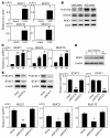
Figure 1. Enhancement of STAT3 activation in CECs by IL-22.
(A) Expression levels of IL-22 in normal colon of WT (n = 9) and inflamed colon of TCRαKO (TCRα) mice (n = 9) and the CD45RB colitis model (n = 4). (B) Expression levels of IL-22 in intestinal biopsy samples from healthy controls (n = 10) and from patients with UC (n = 12) or CD (n = 9) are shown. (C) Expression levels of IL-22 in purified CD4+ T cells and IgM+ cells from inflamed colon of TCRαKO mice are shown. (D) Expression levels of IL-22 in purified CD4+ T cells from inflamed colon of TCRαKO mice and CD45RB model are shown. (E) Expressions of β-actin, IL-22RA1, and IL-10R2 in CECs (red lines) and colonic LP (black lines) of TCRαKO mice. Results represent averages of 4 different experiments ± SEM. Quantitative data relative to β-actin expression are summarized in the 2 right panels. (F and G) Freshly isolated CECs from WT mice were stimulated with various doses of IL-22 for 15 minutes (F). Normal human colonic tissues were stimulated without (cont) or with 10 ng/ml of IL-22 (G). Protein lysates (10 μg) were immunoblotted with anti–phospho-STAT3 (P-STAT3) or anti–phospho–ERK1/2 Abs. After stripping the Abs, the membrane was reprobed with anti-STAT3 or anti-ERK1/2 Abs. The result is representative of 3 individual experiments. (H) Human colonic specimen was stimulated without (top panel) or with IL-22 (bottom panel) for 2 hours and subjected to immunohistochemical analysis using anti–phospho-STAT3 Abs. Original magnification, ×40.
Figure 2
Figure 2. Development of local gene-delivery system that is capable of targeting colon.
(A) A complex of mock or enhanced GFP vector with DOTAP/enhancer reagent was locally microinjected into the proximal part (just below ileocecal junction) of colon of TCRαKO mice thorough the laparotomy approach. The mice were sacrificed 1, 2, or 4 weeks after the microinjection. Frozen sections of the injection site were subjected to fluorescent microscopic analysis for the detection of GFP signals. The result is representative of 6 individual experiments. (B) Adherent cell populations (such as macrophages and fibroblasts) were isolated from the deepithelialized mucosa with GFP vector delivery using a “walk-out” approach. The obtained cells with an adherent ability were subjected to fluorescent microscopic analysis for the detection of GFP signals. Original magnification, ×40.
Figure 3
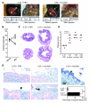
Figure 3. Rapid attenuation of colitis by local IL-22 gene delivery.
(A and B) Complexes of IL-22 secretion or mock vector with DOTAP/enhancer reagent were microinjected into the proximal part of colon of TCRαKO mice with colitis. Expressions of IL-22 in the noninjected distal part and injected proximal part are shown in A. Results represent the averages ± SEM (n = 6–7). **P < 0.005. Protein lysates from the CECs of the proximal part of colon with mock or IL-22 gene delivery were subjected to immunoblot for evaluation of STAT3 activation (B). (CH) Laparotomy was carried out on anesthetized TCRαKO mice (24 weeks of age) to confirm the presence of severe colitis as indicated by a marked enlargement of colonic diameter (before injection). Local gene delivery of IL-22 or mock vector into the proximal part (just below the ileocecal junction) was performed in selected TCRαKO mice (n = 7 each group) that had severe colitis. Mice were sacrificed 2 weeks after microinjection. IL-22 gene delivery attenuated the inflammation at the injection sites; colonic diameter at the injection sites was markedly reduced in TCRαKO mice that received IL-22 (C, right panels) but not mock (C, left panels) vector delivery. Results are summarized in D. Colonic thickness (E) and disease score (F), which were evaluated by histological examination (n = 6–7), are shown. Histology of the proximal colon where gene delivery with mock (left panel) or IL-22 vector (right panel) was received are shown in G. Original magnification, ×10. (H) Percentages of goblet cells among total epithelial cells within the colon where mock (n = 6) or IL-22 (n = 6) vector delivery were received are shown. *P < 0.001.
Figure 4
Figure 4. IL-22–mediated induction of a series of Muc expressions in CECs through STAT3 activation.
(A and B) CECs were freshly isolated from WT mice that had received local gene delivery of mock (white bars, n = 5) or IL-22 secretion (black bars, n = 5) vector into the proximal colon. Expressions of MUCs by CECs are shown in A. Protein lysates from freshly isolated CECs were subjected to immunoblot with anti–phospho-STAT3 (P-STAT3) or anti-MUC1 (MUC1) Abs (B). After stripping the Abs, the membrane was reprobed with anti-STAT3 (STAT3) or actin Abs. (C and D) Average expressions of MUCs by T84 cells stimulated with IL-22 (10 ng/ml) in duplicates of 3 individual experiments are shown in C. Protein lysates were immunoblotted with anti-MUC1 Abs (D). After stripping the Abs, membrane was reprobed with β-actin Abs. (E) T84 cells were transfected with a combination of STAT3 shRNA vectors (pKD-STAT3-v2 and -v3) or STAT1 shRNA vectors (pKD-STAT1-v1 and -v2) using Amaxa, which has been shown to efficiently transfect the gene of interest into cells resistant to typical transfection approaches. Cell line with mock vector transfection was used as control. After stimulation of the shRNA- or mock vector–transfected cells with IL-22 (10 ng/ml), the efficiency of the combined shRNA on the silencing of STAT3 or STAT1 expression was confirmed by Western blot (upper left panels) and real-time PCR analyses (upper right panels). Expression of MUC1, -3, and -13 by shRNA (STAT3 or STAT1) or mock vector–transfected cells stimulated with IL-22 was evaluated by real-time PCR analysis. Data represent the average of 3 individual experiments. **P < 0.005; *P < 0.05.
Figure 7
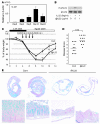
Figure 7. Effect of IL-22BP on the suppression of IL-22 activity in vivo.
(A) Expression levels of IL-22BP (ratio of IL-22BP/β-actin) in the colonic LP at days 0 (normal), 4 (acute phase), and 8 (recovery phase) of DSS colitis are shown. (BF) Local IL-22BP gene delivery was performed in the proximal part of the colon, and the mice were subsequently treated with 3% DSS on the third day after the delivery. DSS treatment was terminated at day 5 after initiation, and mice were sacrificed at day 8. Expression of IL-22BP in the distal (noninjected site) and proximal (injected site) colons of mice with mock or IL-22BP gene delivery is shown (B). CECs were freshly isolated from DSS-treated WT mice that had received local gene delivery of mock or IL-22BP vector in the proximal colon. Protein lysates from the freshly isolated CECs were subjected to immunoblot with anti–phospho-STAT3 Abs (C). After stripping the Abs, the membrane was reprobed with anti-STAT3 Abs (C). The proximal colon of mock (left panel) or IL-22BP (right panel) gene–delivered mice was subjected to Alcian blue staining (D). Significant reduction of goblet cells (blue) is observed in the colon of IL-22BP–delivered mice as compared with that of mock-delivered mice. Ulceration is indicated by arrow. Original magnification, ×4. Averages of goblet cells/field in the noninjected (distal colon) and injected (proximal colon) site are shown in E. Disease score in the proximal colon (injected site) is summarized in F. *P < 0.05; **P < 0.005; ***P < 0.001.
Figure 5
Figure 5. Enhanced mucus production mediates IL-22–induced rapid attenuation of UC-like disease.
(AD) Laparotomy was carried out on anesthetized TCRαKO mice (24 weeks of age) to confirm the presence of colitis as indicated by a marked enlargement of colonic diameter (A, before injection). Local gene delivery of IL-22 vector into the proximal part (just below the ileocecal junction) was performed in the diseased TCRαKO mice. PBS (A, left panels) or mucolytic agent (A, right panels) was continuously administered into the cecal lumen through osmotic pump for 2 weeks. The mice were sacrificed 2 weeks after the microinjection (A, after injection). Numbers in panels indicate the colonic diameter. Summary of change in colonic diameter of 4 mouse groups (n = 4–6) (IL-22 gene delivery plus PBS treatment, black; IL-22 gene delivery plus mucolytic treatment, red; mock gene delivery plus mucolytic treatment, blue; and mock gene delivery plus PBS treatment, green) is shown in B. Histology of the colon from IL-22–gene–delivered TCRαKO mice with PBS (C, left panels) or mucolytic agent (C, right panels) and summarized disease score (D) are shown. (E) Alcian blue staining shows preserved mucus layer (blue liner, arrowhead) along epithelial surface of IL-22–gene–delivered TCRαKO mice (left panels). In contrast, mucolytic treatment impaired the mucus layer formation with significant adhesion of enteric bacteria (arrow; middle panels). Adhesion of enteric bacteria was confirmed by toluidine blue staining (top right panel, arrow). Average (randomly selected 20 fields/each mouse of 4 mice) of thicknesses of bacterial layer attached to epithelial surface is summarized in bottom right panel. **P < 0.001. Original magnification, ×1 (C); ×10 (E, top left panels); ×40 (E, bottom left panels).
Figure 6
Figure 6. Role of IL-22 in the restitution of goblet cells during recovery from acute intestinal injury.
(A) DSS was administered into WT mice for 5 days and the treatment terminated to induce recovery phase. Expression levels of IL-22 (ratio of IL-22/β-actin) in the colonic LP at days 0 (normal), 4 (acute phase), 8, 12 (recovery phases), and 24 are shown (n = 4 each group). (B) CECs were freshly isolated as crypt units from WT mice and stimulated with IL-22 (5 ng/ml) in the presence or absence of anti–IL-22 Abs. Protein lysates (10 μg) from the stimulated CECs were subjected to immunoblot with anti–phospho-STAT3 (P-STAT3) Abs. After stripping Abs, the membrane was reprobed with anti-STAT3 Abs (STAT3). (CE) WT mice were treated with 3.5% DSS in drinking water for 5 days. Repeated i.p. administration of 1.5 mg/injection of anti–IL-22 Abs (@IL22; n = 12) or control Ig (Cont; n = 12) was performed at days 4, 5, 6, and 7 in selected mice that showed similar body weight loss at day 4 as compared with initial body weight. Body weight change (C), “Swiss rolls” (D, upper panels), and high magnification (D, bottom panels) of colons stained with Alcian blue are shown. *P < 0.05; **P < 0.005; ***P < 0.001. Original magnification, ×1 (E, top row); ×20 (E, bottom row). Disease scores are summarized in E.

Comment in

  • IL-22: a two-headed cytokine in IBD?
    Seiderer J, Brand S. Seiderer J, et al. Inflamm Bowel Dis. 2009 Mar;15(3):473-4. doi: 10.1002/ibd.20625. Inflamm Bowel Dis. 2009. PMID: 18668680 No abstract available.

References

    1. Dumoutier L., Louahed J., Renauld J.C. Cloning and characterization of IL-10-related T cell-derived inducible factor (IL-TIF), a novel cytokine structurally related to IL-10 and inducible by IL-9. J. Immunol. 2000;164:1814–1819. - PubMed
    1. Fickenscher H., et al. The interleukin-10 family of cytokines. Trends Immunol. 2002;23:89–96. - PubMed
    1. Pestka S., et al. Interleukin-10 and related cytokines and receptors. Annu. Rev. Immunol. 2004;22:929–979. - PubMed
    1. Liang S.C., et al. Interleukin (IL)-22 and IL-17 are coexpressed by Th17 cells and cooperatively enhance expression of antimicrobial peptides. . J. Exp. Med. 2006;203:2271–2279. - PMC - PubMed
    1. Zheng Y., et al. Interleukin-22, a Th17 cytokine, mediates IL-23-induced dermal inflammation and acanthosis. Nature. 2007;445:648–651. - PubMed

Publication types

MeSH terms

Substances