. 2008 Jul;265(7):843-6.
doi: 10.1007/s00405-007-0531-6. Epub 2008 Jan 3.

Primary temporal bone secretory meningioma presenting as chronic otitis media

Affiliations

Primary temporal bone secretory meningioma presenting as chronic otitis media

T A T Marcelissen et al. Eur Arch Otorhinolaryngol. 2008 Jul.

Abstract

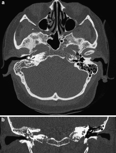
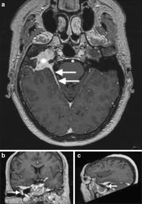
We report an extremely rare case of a secretory meningioma primarily involving the temporal bone. A 56-year old female patient presented to us with a history of a chronic otitis media and unilateral hearing loss. Diagnostic investigations revealed a tumor arising from the temporal bone without signs of intracranial involvement. Histopathological examination showed a meningioma of the secretory type. The tumor was partially resected and serial imaging at follow-up revealed no extension of the tumor. No new symptoms developed 1 year after surgery. Secretory meningioma is a rare meningioma subtype and extracranial presentation in the temporal bone is very unusual. We present the first case of a primary temporal bone secretory meningioma in the otorhinolaryngological literature. As radical as possible surgical excision with serial imaging at follow-up is recommended.


Figures

Fig. 1
Fig. 1
High resolution Computed Tomography (CT) in the axial plane (a) with coronal reconstruction (b) of the petrous bones shows in the right ear middle ear and mastoid cells a soft tissue mass (asterisks). The ossicular chain was intact and not displaced. There was no evidence of destruction of the petrous bone or mastoid
Fig. 2
Fig. 2
Secretory meningeoma with many round pseudo-psammoma bodies (HE, original magnification ×200)
Fig. 3
Fig. 3
a Post-contrast T1–WI MRI of the head. In the middle cranial fossa on the right side, a homogeneous hyper-intense mass on the axial plane can be seen (asterisk), in close contact to the temporal bone. Also thickening and hyper-intense signal intensity of the tentorium (arrows) represent the “dural tail”. b, c On the coronal and sagittal images the extension of the mass into the petrous bone with spread to the epitympanon can be seen (small arrows)

References

    1. O’Reilly RC, Kapadia SB, Kamerer DB. Primary extracranial meningiomas of the temporal bone. Otolaryngol Head Neck Surg. 1998;118(5):690–694. doi: 10.1016/S0194-5998(98)70245-1. - DOI - PubMed
    1. Alguicil-Garcia A, Pettigrew NM, Sima AA. Secretory meningioma: a distinct subtype of meningioma. Am J Surg Pathol. 1986;10:102–111. doi: 10.1097/00000478-198602000-00003. - DOI - PubMed
    1. Kepes JJ. Observation on the formation of psammoma bodies and pseudopsammoma bodies in meningiomas. J Neuropathol Exp Neurol. 1961;34:282–289. doi: 10.1097/00005072-197505000-00005. - DOI - PubMed
    1. Probst-Cousin S, Villagran-Lillo R, Lahl R, Bergmann M, Schmid KW, Gullotta F. Secretory meningioma, clinical, histological and immunohistochemical finding in 31 cases. Cancer. 1997;79:2003–2015. doi: 10.1002/(SICI)1097-0142(19970515)79:10<2003::AID-CNCR23>3.0.CO;2-X. - DOI - PubMed
    1. Gagnon NB, Lavigne F, Mohr G, Guerard MJ, Bouvier G. Extracranial and intracranial meningiomas. J Otolaryngol. 1986;15(6):380–384. - PubMed