. 2008 Mar;123(2):163-76.
doi: 10.1007/s00439-007-0456-6. Epub 2008 Jan 3.

The Opitz syndrome gene product MID1 assembles a microtubule-associated ribonucleoprotein complex

Affiliations

The Opitz syndrome gene product MID1 assembles a microtubule-associated ribonucleoprotein complex

Beatriz Aranda-Orgillés et al. Hum Genet. 2008 Mar.

Abstract

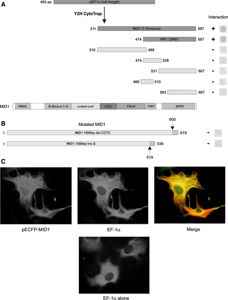
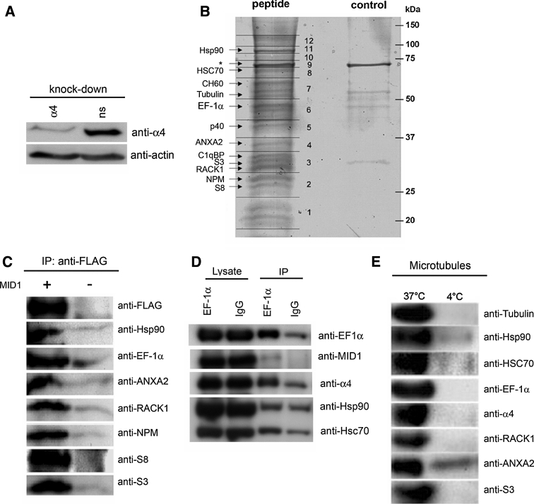
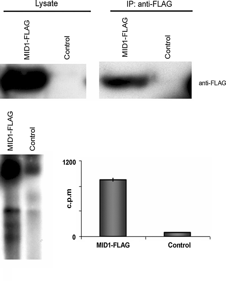
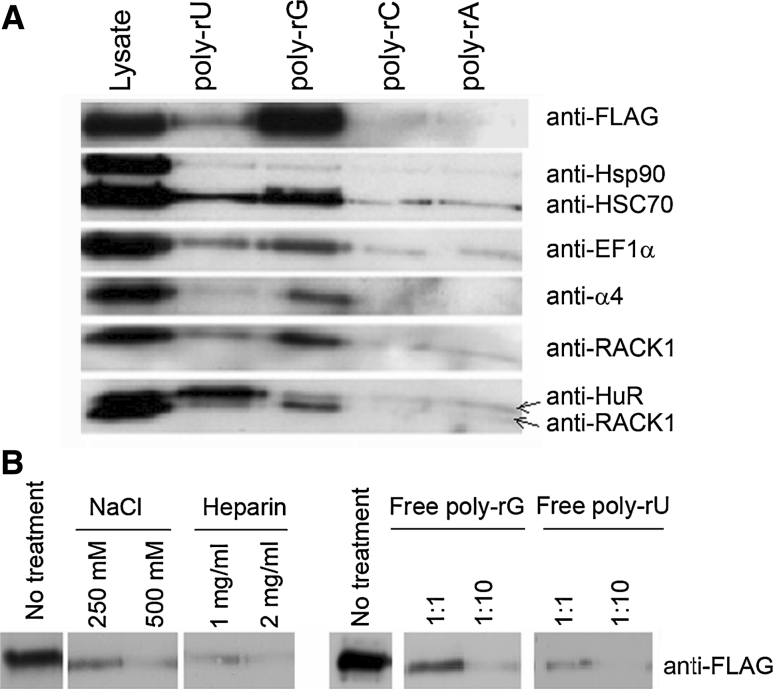
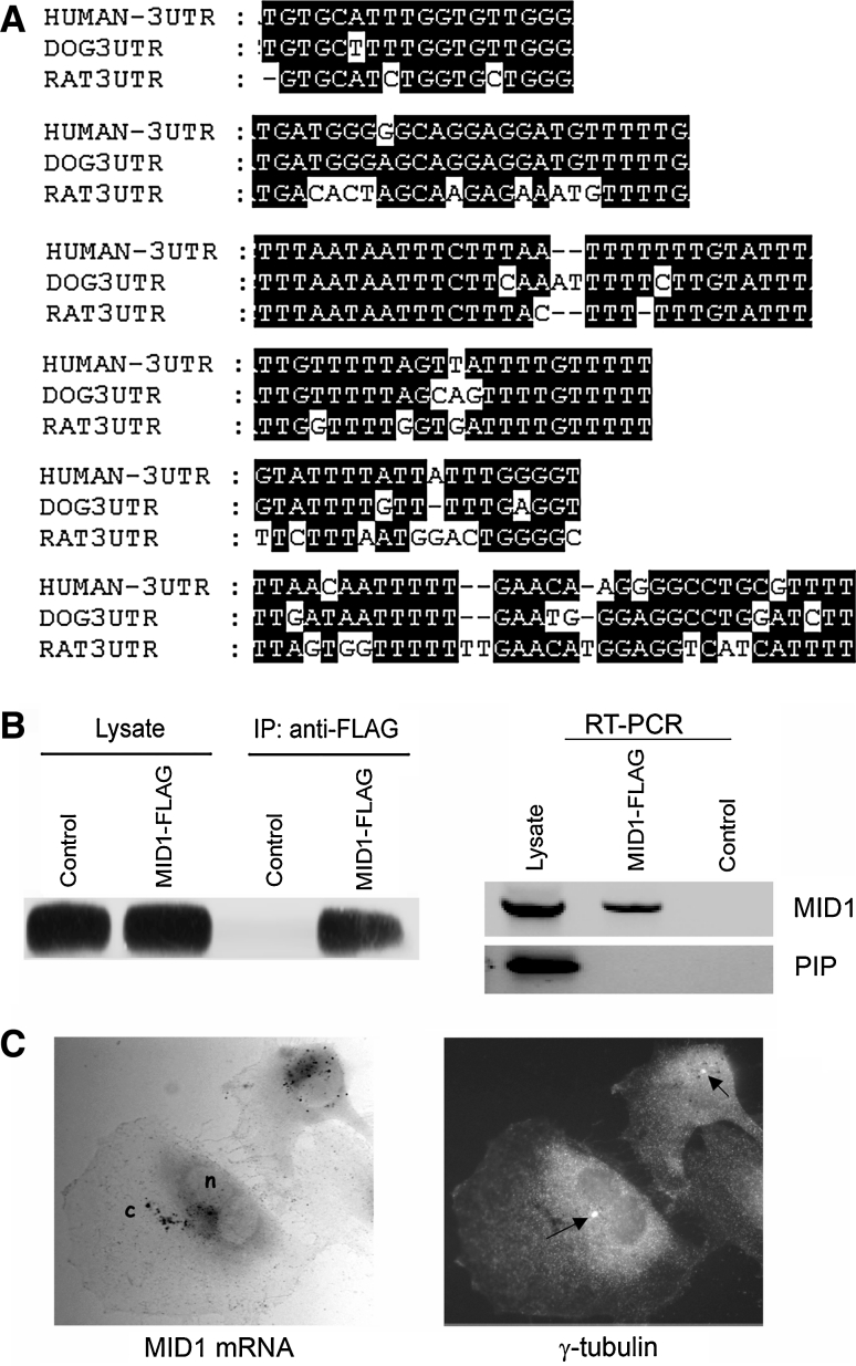
Opitz BBB/G syndrome (OS) is a heterogenous malformation syndrome mainly characterised by hypertelorism and hypospadias. In addition, patients may present with several other defects of the ventral midline such as cleft lip and palate and congenital heart defects. The syndrome-causing gene encodes the X-linked E3 ubiquitin ligase MID1 that mediates ubiquitin-specific modification and degradation of the catalytic subunit of the translation regulator protein phosphatase 2A (PP2A). Here, we show that the MID1 protein also associates with elongation factor 1alpha (EF-1alpha) and several other proteins involved in mRNA transport and translation, including RACK1, Annexin A2, Nucleophosmin and proteins of the small ribosomal subunits. Mutant MID1 proteins as found in OS patients lose the ability to interact with EF-1alpha. The composition of the MID1 protein complex was determined by several independent methods: (1) yeast two-hybrid screening and (2) immunofluorescence, (3) a biochemical approach involving affinity purification of the complex, (4) co-fractionation in a microtubule assembly assay and (5) immunoprecipitation. Moreover, we show that the cytoskeleton-bound MID1/translation factor complex specifically associates with G- and U-rich RNAs and incorporates MID1 mRNA, thus forming a microtubule-associated ribonucleoprotein (RNP) complex. Our data suggest a novel function of the OS gene product in directing translational control to the cytoskeleton. The dysfunction of this mechanism would lead to malfunction of microtubule-associated protein translation and to the development of OS.


Figures

Fig. 1
Fig. 1
a EF-1α interacts with the MID1 protein. a Deletion mapping of the EF-1α binding site on the MID1 protein in a yeast two-hybrid assay. While two C-terminal peptides (aa 311- aa 667 and aa 474- aa 667) show clear binding to EF-1α full-length protein, no binding was seen with any of the shorter constructs, as indicated on the right panel by the appearance or lack of yeast growth, respectively. b Yeast two-hybrid interaction assay between EF-1α full-length and two mutated MID1 variants as found in Opitz syndrome patients. Both MID1 mutations result in a frame shift and a truncation of the resulting MID1 proteins. The upper MID1 variant harbours a 4 bp deletion at position 1800 and the lower one a G-insertion at position 1558 resulting in a wrong reading frame after amino acid 600 and 519, respectively (arrows), the hatched regions indicate the amino acid sequence corresponding to the wrong reading frame until the first stop codon is reached. On the right panel the respective parts of the yeast plates are shown; they show no growth and hence no interaction. c Immunofluorescence microscopy of COS-7 cells transiently transfected with ECFP-MID1 and EF-1α (upper panel) and EF-1α alone (lower panel). The distribution of EF-1α when expressed alone shows a diffuse cytoplasmic staining patter, whereas co-expressed with ECFP-MID1 it co-localises with ECFP-MID1 at the microtubules
Fig. 2
Fig. 2
a Knockdown of α4 protein in HeLa cells- α4 protein in HeLa cell lysates after transfection with a specific α4 RNAi oligonucleotide (α4, first lane) or a non-silencing oligonucleotide (ns, second lane) are shown. Detection of actin was used as loading control. b SDS-gel stained with colloidal Coomassie showing the proteins eluted from streptavidin beads coupled to a biotinylated 44 aa α4 peptide (left lane) or from unmodified beads (right lane). Differential bands were excised and analysed by mass spectrometry. Protein identities are given. The asterisk indicates the position of the MID1 protein band, which is masked by an abundant E. coli band derived protein band contaminating the eluting peptide (also present in the control). c Co-immunoprecipitation of FLAG-MID1 with different components of the complex. HeLa cell lysates overexpressing FLAG-MID1 were immunoprecipitated with anti-FLAG antibody (left lane). Western blots were incubated with specific antibodies against the respective proteins. HeLa cell lysates without FLAG-MID1 overexpression were used as background control (right lane). d Co-immunoprecipitation of endogenous EF-1α with MID1, α4, Hsp90 and Hsc70. HeLa cell lysates were immunoprecipitated with anti-EF1α (lanes 1 and 3) or with unspecific IgGs (lanes 2 and 4). Input lysates (lanes 1 and 2) and immunoprecipitates (lanes 3 and 4) were loaded on an SDS-Page and analysed with anti-EF-1α, anti-MID1, anti-α(, anti-Hsp90 and anti-Hsc70 antibodies. e Microtubule-association of the complex partners. Pellets from microtubule-assembly experiments at 37°C (left lane) and 4°C (right lane) were dissolved and loaded on a Western blot and detected with the respective antibodies
Fig. 3
Fig. 3
The MID1 protein complex associates with RNA. Cytosol of HeLa cells with (FLAG-MID1) and without (control) FLAG-MID1 overexpression were immunoprecipitated using an anti-FLAG antibody. Immunoblots of lysates (upper left panel) and immunoprecipitates (upper right panel) probed with an anti-FLAG antibody are shown. RNA was isolated from the samples, labelled with [5´-32P]pCp and analysed on an agarose gel (lower left panel) and in a scintillation counter (lower right panel)
Fig. 4
Fig. 4
Association of FLAG-MID1 and some of the complex partners with poly-ribonucleotides. a Lysates from FLAG-MID1 overexpressing HeLa cells were incubated with immobilised poly-rU (lane 2), poly-rG (lane 3), poly-rC (lane 4) or poly-rA (lane 5), washed and boiled at 95°C. Lysate (lane 1) and eluted fractions were immunoblotted and analysed with the respective antibodies. Poly-rU binding HuR protein was used as control. b Influence of free poly-rG and poly-rU competitors and increasing salt or heparin concentrations on the FLAG-MID1/poly-rG interaction
Fig. 5
Fig. 5
The MID1 complex assembles its own mRNA. a G and U-rich sequences present in the MID1 3′UTR that show a high interspecies conservation. Positions 3′ to the translational stop codon are (from top to bottom) 3101, 3172, 185, 1502, 2441 and 2644 bp. Accession number NM_000381. b HeLa cells were transfected with FLAG-MID1 and subjected to RNP coimmunoprecipitation. Subsequently, RNA was isolated from the precipitation and subjected for RT-PCR using MID1 and PIP-specific primers. Immunoprecipitation was done with mouse IgG as a negative control. c The MID1 mRNA localises to the centrosomal region. The endogenous MID1 mRNA was detected in U373 cells by in situ hybridisation using a pool of six different digoxigenin-labelled oligonucleotides specific for the MID1 sequence and alkaline phosphatase-linked anti-digoxigenin antibody with NBT/BCIP as substrate. The centrosome was co-stained by a mouse anti-γ-tubulin antibody and an FITC-linked anti-mouse antibody (see arrows). n nucleus, c cytoplasm

References

    1. Angenstein F, Evans AM, Settlage RE, Moran ST, Ling S-C, Klintsova AY, Shabanowitz J, Hunt DF, Greenough WT. A Receptor for Activated C Kinase Is Part of Messenger Ribonucleoprotein Complexes Associated with PolyA-mRNAs in Neurons. J Neurosci. 2002;22:8827–8837. - PMC - PubMed
    1. Balch C, Dedman JR. Annexins II and V inhibit cell migration. Exp Cell Res. 1997;237:259–263. doi: 10.1006/excr.1997.3817. - DOI - PubMed
    1. Bashirullah A, Cooperstock RL, Lipshitz HD. RNA localization in development. Annu Rev Biochem. 1998;67:335–394. doi: 10.1146/annurev.biochem.67.1.335. - DOI - PubMed
    1. Bassell GJ, Singer RH. Neuronal RNA localization and the cytoskeleton. Results Probl Cell Differ. 2001;34:41–56. - PubMed
    1. Bertrand E, Chartrand P, Schaefer M, Shenoy SM, Singer RH, Long RM. Localization of ASH1 mRNA particles in living yeast. Mol Cell. 1998;2:437–445. doi: 10.1016/S1097-2765(00)80143-4. - DOI - PubMed

Publication types

MeSH terms

LinkOut - more resources