. 2008 Mar;17(3):361-372.
doi: 10.1007/s00586-007-0566-9. Epub 2008 Jan 3.

A single posterior approach for multilevel modified vertebral column resection in adults with severe rigid congenital kyphoscoliosis: a retrospective study of 13 cases

Affiliations

A single posterior approach for multilevel modified vertebral column resection in adults with severe rigid congenital kyphoscoliosis: a retrospective study of 13 cases

Yan Wang et al. Eur Spine J. 2008 Mar.

Abstract

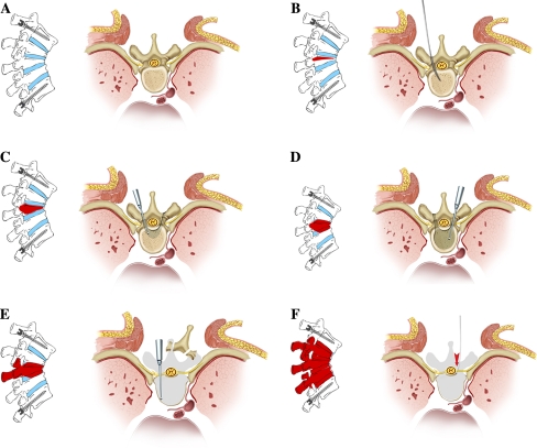
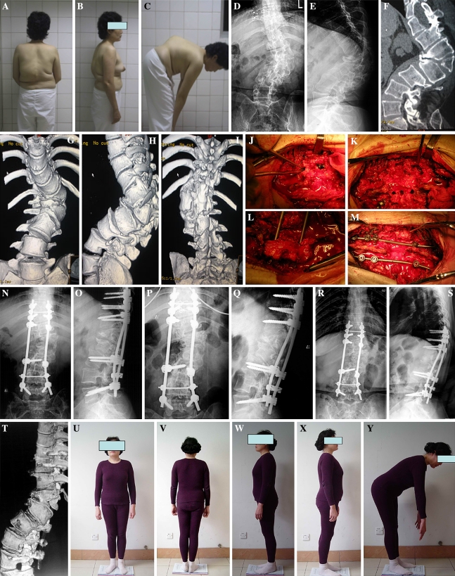
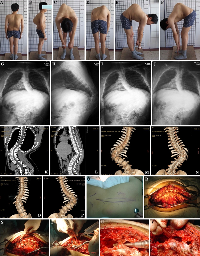
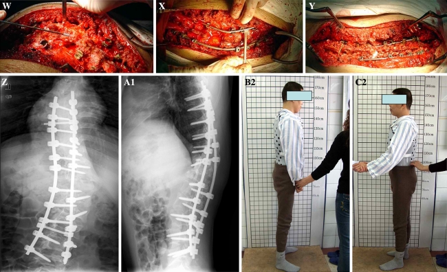
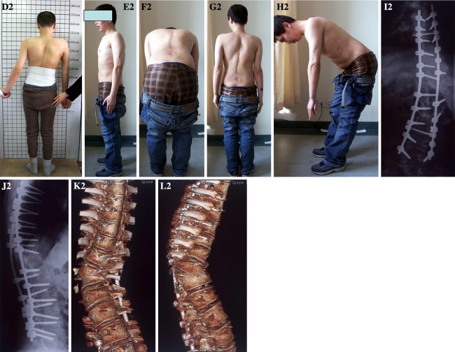
We report a multilevel modified vertebral column resection (MVCR) through a single posterior approach and clinical outcomes for treatment of severe congenital rigid kyphoscoliosis in adults. Transpedicular eggshell osteotomies and vertebral column resection are two techniques for the surgical treatment of rigid severe spine deformities. The authors developed a new technique combining the two surgical methods as a MVCR, through a single posterior approach, for surgical treatment of severe congenital rigid kyphoscoliosis in adults. Thirteen adult patients with severe rigid congenital kyphoscoliosis deformity were treated by a single posterior approach using a MVCR technique. The surgery processes included a one-stage posterior transpedicular eggshell technique first, and then expanded the eggshell technique to adjacent intervertebra space through abrasive reduction of the vertebral cortices from inside out. All posterior vertebral elements were removed including the cortical vertebral bone around the neural canal. Range of resection of the vertebral column at the apex of the deformity included apical vertebra and both cephalic and/or caudal adjacent wedged vertebrae. Totally, 32 vertebrae had been removed in 13 patients, with 2.42 vertebrae being removed on average in each case. The average fusion extent was 7.69 vertebrae. Mean operation time was 266 min with average blood loss of 2,411.54 ml during operation. Patients were followed up for an average duration of 2.54 years. Deformity correction was 59% in the coronal plane (from 79.7 degrees to 32.4 degrees ) postoperatively and 33.7 degrees (57% correction) at 2 years follow-up. In the sagittal plane, correction was from preoperative 85.9 degrees to 27.5 degrees immediately after operation, and 32.0 degrees at 2 years follow-up. Postoperative pain was reduced from preoperative 1.77 to 0.54 at 2 years follow-up in visual analog scale. SRS-24 scale was from 38.2 preoperatively to 76.9 at 2 years follow-up postoperative. Complications were encountered in four patients (30.7%) with transient neurology that spontaneously improved without further treatment within 3 months. MVCR technique through a single posterior approach is an effective procedure for the surgical treatment of severe congenital rigid kyphoscoliosis in adults.


Figures

Fig. 1
Fig. 1
Diagram illustration of the MVCR procedure. a Pedicle screws were inserted into the vertebrae; a stabilizing rod was preset on the concaved side. b A probe was used to determine the entry point and depth in the dissecting segment. c After enlarging the entry with a curette, a 5-mm burr was used to perform regular “eggshell” technique, and during this process, the inner wall of the pedicle was kept as intact as possible. d The entry was further enlarged both cephalically and caudally to penetrate the corresponding walls, and small amount of bony wall was kept to protect the nerve roots. e When the end-plates of the upper and lower intervertebral space were exposed, a Kerrison rongeur or burr was used to abrade the anterior and lateral walls of the vertebral body, which was then collapsed under pressure laterally to expose the posterior walls further. f Then a similar vertebral body resection was advanced to cross the midline in an abrasive way with the burr. Adjacent upper and lower end-plates as well as intervertebral discs were removed with a curette
Fig. 2
Fig. 2
A 43-year-old woman who presented with hemivertebra kyphoscoliosis deformity with dysplasia of several vertebrae adjacent to the apical vertebra. a–c Preoperative outlook. d, e Radiographic evaluation found that the apical hemivertebra of kyphoscoliosis was L1; the curve in coronal and sagittal plane was 75.4° and 84.1°, respectively. f–i Computerized tomography and 3-D reconstruction images revealed that the apical vertebra and its adjacent vertebrae of T12 and L2 had severe wedged deformity on anterior, posterior, and lateral views having a local kyphosis deformity of 95.2°, which was far more severe than X-ray measurement. j–m MVCR surgical procedure was carried out to resect the vertebrae of T12, L1, and L2, with posterior pedicle screw fixation, vertebrae compression, and fusion with bone chip autograft. n, o The deformity was corrected to 36.3° and 12.2° in the coronal and sagittal plane, respectively, as shown in the corresponding radiographs. p, q 6-month follow-up. r, s 2-year follow-up X-ray measurement: her deformity was 36.4° and 25.6° in the coronal and sagittal plane, respectively, and fusion can be found both in X-ray and 3-D reconstruction images. t 3-D reconstruction images. u–y 2-year follow-up outlook image
Fig. 3
Fig. 3
a–f A 30-year-old man who presented with hemivertebra (T11) kyphoscoliosis deformity with dysplasia of several vertebrae adjacent to the apical vertebra. g–j His preoperative coronal and sagittal deformities were 110.8° and 79.3°, respectively. k–p Computerized tomography and 3-D reconstruction images revealed that the apical vertebra and its adjacent vertebrae of T10 and T12 had severe wedged deformity on anterior, posterior, and lateral views. q–y He was treated with three-level MVCR to resect the vertebrae of T10, T11, and T12, with posterior pedicle screw fixation, vertebrae compression, and fusion with bone chip autograft, and 12 segments were fused. z–a1 Right after operation, his deformity correction in coronal and sagittal plane was 34.2° and 35.5°, respectively. b2–d2 The patient outlook image 2 weeks after operation; he needed other people’s help to stand up because of his recent recovery from temporary paraplegina postoperatively. e2–h2 The patient outlook image 2 years after operation. i2–l2 Two-year follow-up X-ray measurement: his deformity was 33.1° and 39.3° in the coronal and sagittal plane, respectively, and fusion can be found both in X-ray and 3-D reconstruction images
Fig. 3
Fig. 3
a–f A 30-year-old man who presented with hemivertebra (T11) kyphoscoliosis deformity with dysplasia of several vertebrae adjacent to the apical vertebra. g–j His preoperative coronal and sagittal deformities were 110.8° and 79.3°, respectively. k–p Computerized tomography and 3-D reconstruction images revealed that the apical vertebra and its adjacent vertebrae of T10 and T12 had severe wedged deformity on anterior, posterior, and lateral views. q–y He was treated with three-level MVCR to resect the vertebrae of T10, T11, and T12, with posterior pedicle screw fixation, vertebrae compression, and fusion with bone chip autograft, and 12 segments were fused. z–a1 Right after operation, his deformity correction in coronal and sagittal plane was 34.2° and 35.5°, respectively. b2–d2 The patient outlook image 2 weeks after operation; he needed other people’s help to stand up because of his recent recovery from temporary paraplegina postoperatively. e2–h2 The patient outlook image 2 years after operation. i2–l2 Two-year follow-up X-ray measurement: his deformity was 33.1° and 39.3° in the coronal and sagittal plane, respectively, and fusion can be found both in X-ray and 3-D reconstruction images

References

    1. Baba H, Maezawa Y, Kamitani K, et al. Osteoporotic vertebral collapse with late neurological complications. Paraplegia. 1995;33:281–289. - PubMed
    1. Boachie-Adjei O, Ferguson JA, Pigeon RG, Peskin MR. Transpedicular lumbar wedge resection osteotomy for fixed sagittal imbalance: surgical technique and early results. Spine. 2006;31(4):485–492. doi: 10.1097/01.brs.0000199893.71141.59. - DOI - PubMed
    1. Bradford DS, Boachie-Adjei O. One-stage anterior and posterior hemivertebral resection and arthrodesis for congenital scoliosis. J Bone Joint Surg Am. 1990;72:536–540. - PubMed
    1. Bradford DS, Tribus CB. Vertebral column resection for the treatment of rigid coronal decompensation. Spine. 1997;22(14):1590–1599. doi: 10.1097/00007632-199707150-00013. - DOI - PubMed
    1. Bridwell KH, Lewis SJ, Edwards C, Lenke LG, Iffrig TM, Berra A, Baldus C, Blanke K. Complications and outcomes of pedicle subtraction osteotomies for fixed sagittal imbalance. Spine. 2003;28(18):2093–2101. doi: 10.1097/01.BRS.0000090891.60232.70. - DOI - PubMed

MeSH terms