Review
. 2008 Apr;36(4):554-62.
doi: 10.1007/s10439-007-9426-3. Epub 2008 Jan 3.

Effects of disturbed flow on endothelial cells

Affiliations
Review

Effects of disturbed flow on endothelial cells

Shu Chien. Ann Biomed Eng. 2008 Apr.

Erratum in

  • Ann Biomed Eng. 2010 Mar;38(3):1258

Abstract

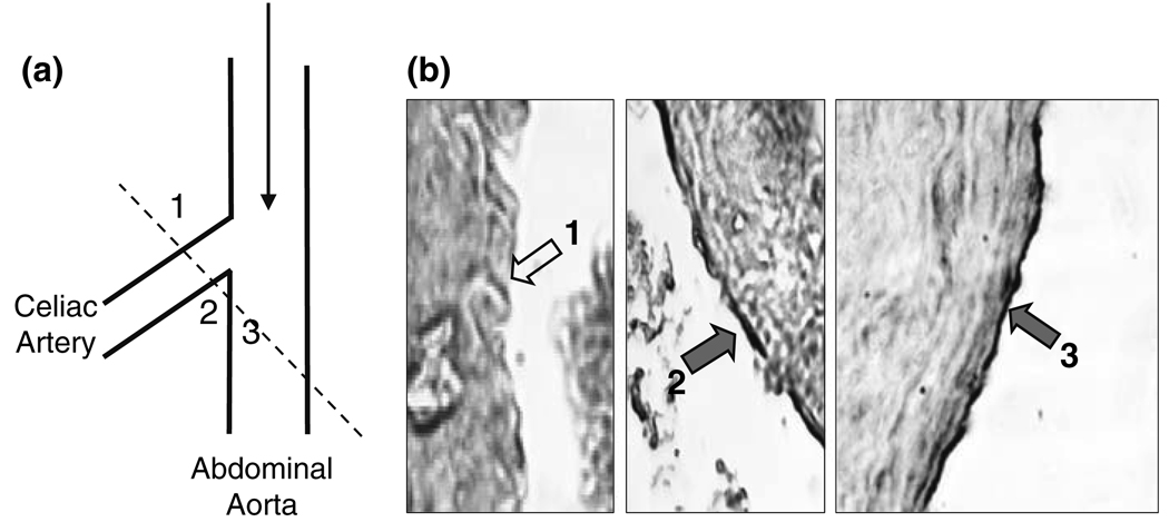
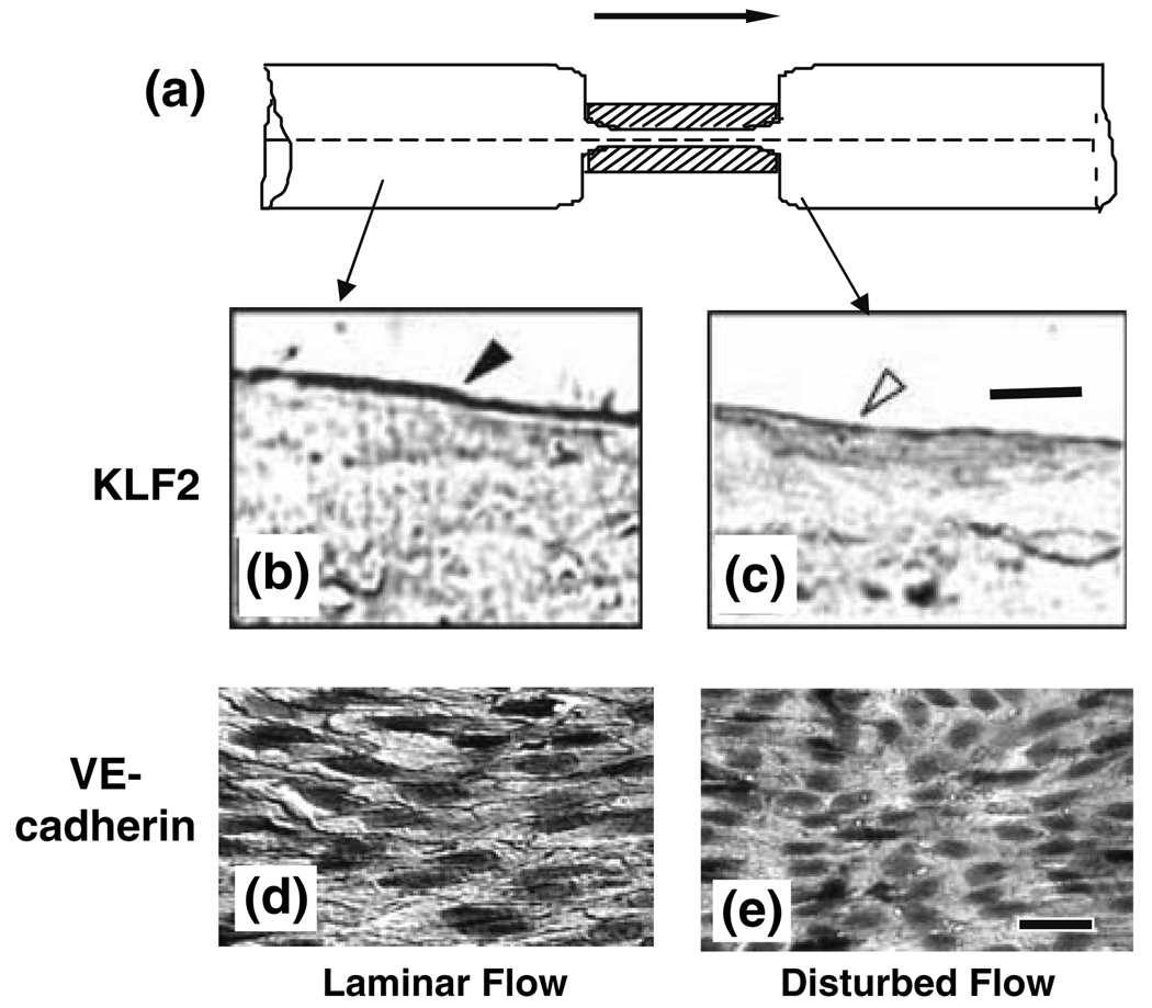
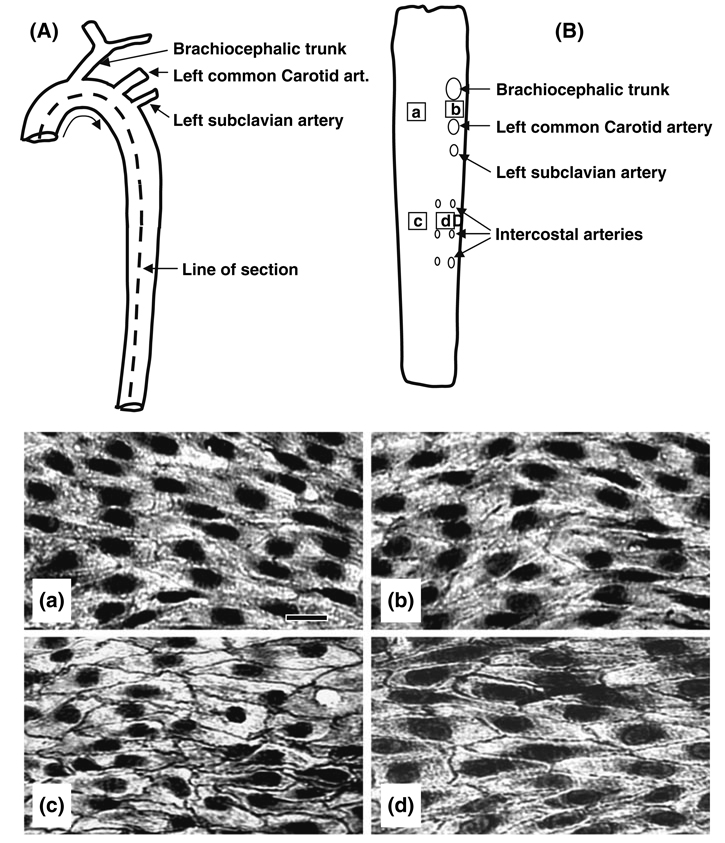
Vascular endothelial cells (ECs) play significant roles in regulating circulatory functions. The shear stress resulting from blood flow modulates EC functions by activating mechano-sensors, signaling pathways, and gene and protein expressions. Shear stress with a clear direction resulting form pulsatile or steady flow causes only transient activation of pro-inflammatory and proliferative pathways, which become down-regulated when such directed shearing is sustained. In contrast, shear flow without a definitive direction (e.g., disturbed flow in regions of complex geometry) causes sustained molecular signaling of pro-inflammatory and proliferative pathways. The EC responses to shear flows with a clear direction involve the remodeling of EC structure to maintain vascular homeostasis and are athero-protective. Such regulatory mechanism does not operate effectively when the flow pattern is disturbed. Therefore, the branch points and other regions of the arterial tree with a complex geometry are prone to atherogenesis, whereas the straight part of the arterial tree is generally spared. Understanding of the EC responses to different flow patters helps to elucidate the mechanism of the region-specific localization of atherosclerosis in the arterial system.


Figures

FIGURE 1
FIGURE 1
Side views of rectangular flow chamber (a) and step flow chamber (b). In both bases, the shear flow can be either steady without oscillations or superimposed with sinusoidal oscillation by using the oscillatory pump. The EC monolayer is shown only in (a); the inset in (a) shows an enlarged view of two ECs and the velocity gradient due to the applied shear stress. In (b), the flow direction in zone b is opposite to the incoming flow and to that in zone d, where forward flow is re-established. Zone c is the reattachment zone where the flow fluctuates between the forward and backward directions, with a low shear stress but large shear stress gradient; this is the area of disturbed flow. From Chien.
FIGURE 2
FIGURE 2
Flow-pattern-specific regulation of KLF2 gene expression in human umbilical vein endothelial cells. (a) Pulsatile flow with a sinusoidal oscillatory shear stress of 1.2 ± 0.4 Pa. (b) Reciprocating shear stress with a very low mean shear stress of 0.05 Pa (to maintain nutrient delivery) and a sinusoidal oscillation of ±0.4 Pa. (c) KLF2 mRNA levels of confluent ECs subjected to pulsatile shear at 1.2 ± 0.4 Pa (PS) for 0, 1, 4, 12, and 24 h. Note the sustain elevation of KLF-2 over the 24 h of study. (d) KLF2 mRNA levels of confluent ECs subjected to reciprocating shear at 0.05 ± 0.4 Pa (RS) for 0, 1, 4, 12, and 24 h. Note the transient elevation of KLF-2 followed by a decline to a level below control at 24 h. Based on Wang et al.
FIGURE 3
FIGURE 3
VE-cadherin staining in endothelial cells exposed to disturbed and laminar flows in vitro. Confluent monolayers of BAECs were kept as controls or subjected to disturbed and laminar flows in a step flow chamber. Staining of VE-cadherin was observed by confocal microscopy. VE-cadherin staining was continuously distributed around the entire periphery of cells under static condition (a). After a 24-h exposure to the disturbed flow, the VE-cadherin distribution became discontinuous (b), whereas 24-h exposure to laminar flow resulted in a continuous distribution of VE-cadherin (c), as in the static condition. Images shown here are representative results from 3 separate experiments. Bar in a = 30 µm. From Miao et al.
FIGURE 4
FIGURE 4
Effects of flow pattern on the activation of SREBP1 in a step flow chamber. (a) Shows side view of the step flow chamber in which confluent cultured BAECs are subjected to different flow patterns. After shearing for 1 or 12 h, the cells were fixed and immunostained for SREBP1. While disturbed flow induced a sustained activation of SREBP1, as indicated by its translocation into the nuclei (b, Lower left), laminar flow activated SREBP1 in a transient manner, as evidenced by the lack of nuclear staining of SREBP1 at 12 h (b, lower right). Modified from Liu et al.
FIGURE 5
FIGURE 5
KLF2 Expression in ECs at abdominal-celiac branch point. (a) Drawing to show the branch point of celiac artery off the abdominal aorta. The broken line shows the section through which the samples were taken for immunohistochemical examination of KLF2 expression in b. (b) Cross section of the branch area with areas 1, 2, and 3 shown in the three pictures. The expression of KLF2 was high and continuous on ECs of the abdominal aorta (labeled 3) and the medial aspect (labeled 2) of the celiac branch, but virtually absent on the lateral aspect (labeled 1) of the branch. These results are representative of three independent experiments. In (b), the short filled arrows (2 and 3) indicate the positive staining for KLF2 protein, and the open arrow (1) shows the absence of KLF2 protein in the endothelium. Modified from Wang et al.
FIGURE 6
FIGURE 6
(a) Side view of in the rat abdominal aorta with a local stenosis created by using a U-shaped titanium clip, with the aim of studying the effects of flow patterns on KLF2 expression and VE-cadherin distribution in vivo. Four weeks after the creation of stenosis, the vessel was harvested, fixed, embedded, sectioned longitudinally, and subjected to immunohistochemistry staining with anti-KLF2 (b and c) or anti-VE-cadherin (d and e). Blood flow is from left to right, as indicated by the arrow. (b) Endothelium in a laminar flow region 5 mm upstream of the stenosis site shows strong KLF2 staining (closed arrowhead). The downstream laminar flow region (not shown) has the same strong staining. (c) the poststenotic site distal to the clip, which has disturbed flow pattern, shows a lack of KLF2 staining (open arrowhead). Bar in c = 10 µm. (d) In the laminar flow region 5 mm upstream (as well as 5 mm downstream) to the clip site, VE-cadherin is highly expressed at endothelial cell borders. (e) No detectable VE-cadherin is found at cell borders in the disturbed flow region immediately downstream to the constriction. Bar in e = 30 µm. Based on Wang et al. and Miao et al.
FIGURE 7
FIGURE 7
Schematic diagrams of the rat thoracic aorta, showing its overall anatomy (A) and its luminal surface after having been cut open and pinned flat (B), the labels a, b, c and d in B indicate the areas from which the four pictures (a, b, c, and d) for VE-cadherin staining were taken by en face staining with an anti-VE-cadherin antibody. The VE-cadherin staining at endothelial cell borders was weak and discontinuous in the aortic arch (a: inferior and b: superior aspects), where disturbed flow dominates, and was much stronger and continuous in the descending thoracic aorta with laminar flow (c: dorsal and d: ventral aspects). Bar in a = 30 µm. Based on Miao et al.

References

    1. Chen YL, Jan KM, Lin HS, Chien S. Ultrastructural studies on macromolecular permeability in relation to endothelial cell turnover. Atherosclerosis. 1995;118:89–104. - PubMed
    1. Chien S. Molecular and mechanical bases of focal lipid accumulation in arterial wall. Prog. Biophys. Mol. Biol. 2003;83:131–151. - PubMed
    1. Chien S. Molecular basis of rheological modulation of endothelial functions: importance of stress direction. Biorheology. 2006;43:95–116. - PubMed
    1. Chien S. Mechanotransduction and endothelial cell homeostasis: the wisdom of the cell. Am. J. Physiol. Heart Circ. Physiol. 2007;292:H1209–H1224. - PubMed
    1. Chien S, Lin SJ, Weinbaum S, Lee MM, Jan KM. The role of arterial endothelial cell mitosis in macromolecular permeability. Adv. Exp. Med. Biol. 1988;242:59–73. - PubMed

Publication types

LinkOut - more resources