. 2008 Feb;11(1):97-112.
doi: 10.1089/rej.2007.0587.

Elastin haploinsufficiency induces alternative aging processes in the aorta

Affiliations

Elastin haploinsufficiency induces alternative aging processes in the aorta

Mylène Pezet et al. Rejuvenation Res. 2008 Feb.

Abstract

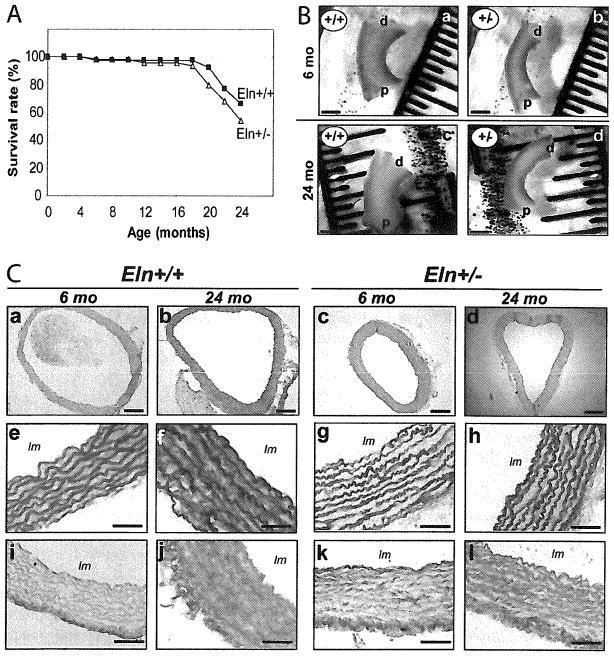
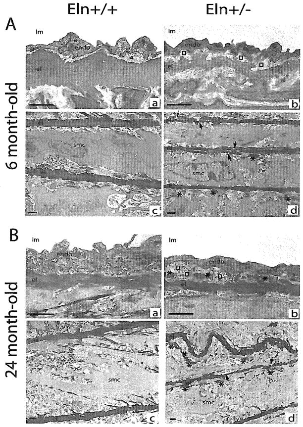
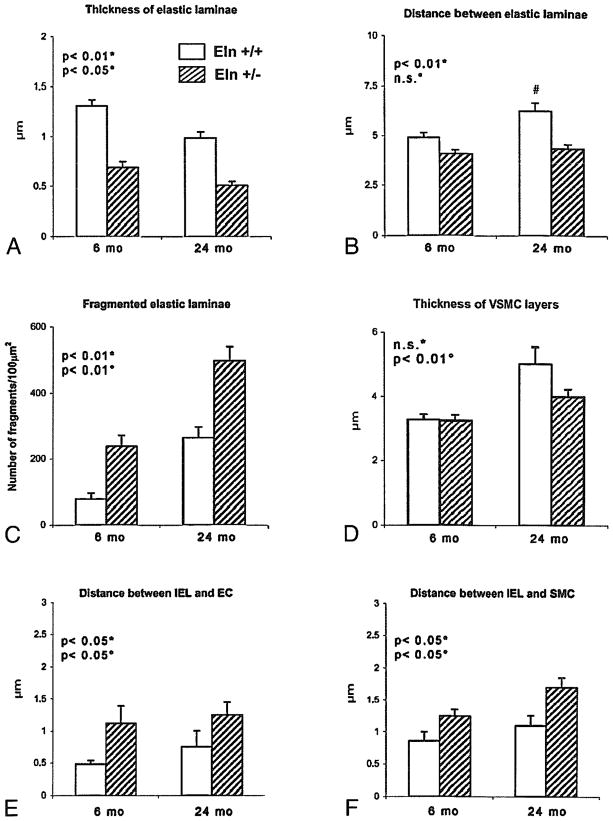
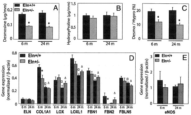
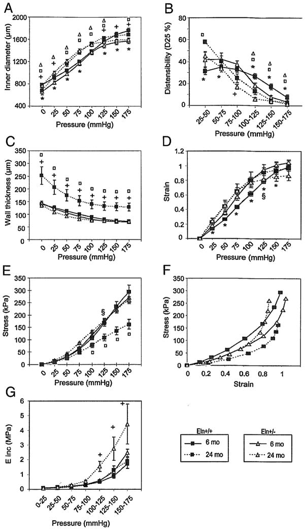
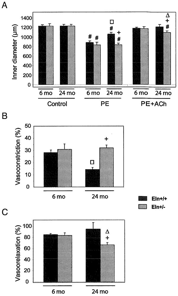
Elastin, the main component of elastic fibers, is synthesized only in early life and provides the blood vessels with their elastic properties. With aging, elastin is progressively degraded, leading to arterial enlargement, stiffening, and dysfunction. Also, elastin is a key regulator of vascular smooth muscle cell proliferation and migration during development since heterozygous mutations in its gene (Eln) are responsible for a severe obstructive vascular disease, supravalvular aortic stenosis, isolated or associated to Williams syndrome. Here, we have studied whether early elastin synthesis could also influence the aging processes, by comparing the structure and function of ascending aorta from 6- and 24-month-old Eln+/- and Eln+/+ mice. Eln+/- animals have high blood pressure and arteries with smaller diameters and more rigid walls containing additional although thinner elastic lamellas. Nevertheless, longevity of these animals is unaffected. In young adult Eln+/- mice, some features resemble vascular aging of wild-type animals: cardiac hypertrophy, loss of elasticity of the arterial wall through enhanced fragmentation of the elastic fibers, and extracellular matrix accumulation in the aortic wall, in particular in the intima. In Eln+/- animals, we also observed an age-dependent alteration of endothelial vasorelaxant function. On the contrary, Eln+/- mice were protected from several classical consequences of aging visible in aged Eln+/+ mice, such as arterial wall thickening and alteration of alpha(1)-adrenoceptor-mediated vasoconstriction. Our results suggest that early elastin expression and organization modify arterial aging through their impact on both vascular cell physiology and structure and mechanics of blood vessels.


Figures

FIG. 1
FIG. 1
Effect of aging and genotype on the survival rate (A), length of the ascending aorta of 6- and 24-month-old Eln+/+ and Eln+/− mice (B), and histological examination of paraffin-embedded cross-sections of the ascending aorta of 6- and 24-month-old Eln+/+ and Eln+/− mice (C). (a–d) eosin/hematoxylin; (e–h) Weigert; and (I–l) picrosirius red stainings (For color version, see <www.liebertonline.com/rej>.). Bar sizes: 200 μm (a–d) and 50 μm (e–l). In each group, n = 39–50 (A) and n = 4–5 (B). lm, lumen; d, distal; p, proximal.
FIG. 2
FIG. 2
Transmission electron microscopy images of the ascending aorta in mice aged of 6 (A) and 24 months (B). (a,c) Eln+/+; (b,d) Eln+/−; (a,b) intima; (c,d) media. Arrows, disruptions of the elastic lamella; asterisks, elastin deposit in margin of elastic lamellas; squares, subendothelial accumulation of extracellular matrix; endo, endothelium; el, elastic lamella; lm: lumen; smc, smooth muscle cell. Bar size: 1 μm.
FIG. 3
FIG. 3
Morphometric analysis of the transmission electron microscopy images of the ascending aorta in Eln+/+ and Eln+/− mice aged of 6 (adult) and 24 months (old). Thickness of the elastic laminae (A), distance between successive elastic laminae (B), fragmentation of elastic laminae (C), thickness of VSMC layer (D), distance between the internal elastic lamina (IEL) and endothelial cells (EC) (E) or smooth muscle cells (SMC) (F). Number of measurements performed on different sections from several animals in each group: 89 (A), 102 (B), 20 (C), 57 (D), 64 (E), 75 (F). #significant difference compared to adult animals of matching genotype. Two-way ANOVA: *general effect of genotype; °general effect of age; n.s., not significant.
FIG. 4
FIG. 4
Desmosine and hydroxyproline contents and ratios (A–C), and gene expressions quantified by real-time PCR (D and E) in the ascending aorta of 6- and 24-month-old Eln+/+ and Eln+/− mice. ELN, tropoelastin; COL1A1, α1 chain of collagen-I; LOX, lysyl-oxidase; LOXL1, lysyl-oxidase-like-1; FBN1, fibrillin-1; FBN2, fibrillin-2; FBLN5, fibulin-5; eNOS, endothelial nitric oxide synthase.*, , Significant difference between Eln+/+ and Eln+/− mice of matching age, between 6- and 24-month-old Eln+/+ mice, and between 6- and 24-month-old Eln+/− mice, respectively. n = 7–11 in each group.
FIG. 5
FIG. 5
Diameter pressure curves and derived mechanical parameters of the ascending aorta of 6- and 24-month-old Eln+/+ and Eln+/− mice. *, +Significant difference between 6-month-old Eln+/+ and Eln+/− mice or between 24 month-old Eln+/+ and Eln+/− mice, respectively. , Significant difference between 6 month-old and 24 month-old values for Eln+/+ and Eln+/− mice, respectively. §Significant difference between Eln+/+ and Eln+/− of matching age at their respective mean arterial pressure. n = 5–10 in each group.
FIG. 6
FIG. 6
Reactivity to vasoactive agents of the ascending aorta of 6- and 24-month-old Eln+/+ and Eln+/− mice. (A) 5 μmol/L phenylephrine (PE) or 5 μmol/L phenylephrine + 5 μmol/L acetylcholine (PE + Ach). PE-induced vasoconstriction is also represented as the decrease in inner diameter, in percent of the control diameter (B), and Ach-induced vasodilation is represented as the reversal of the PE-induced vasoconstriction, in percent (C). #Significant difference between control and post-treatment values at same age and genotype. +, , ,Significant difference for the same treatment between Eln+/+ and Eln+/− of matching ages, between 6- and 24-month-old Eln+/+, and between 6- and 24-month-old Eln+/− mice, respectively. n = 5–9 in each animal group.

References

    1. Rosenbloom J, Abrams WR, Mecham R. Extracellular matrix 4: the elastic fiber. Faseb J. 1993;7:1208–1218. - PubMed
    1. Wolinsky H, Glagov S. A lamellar unit of aortic medial structure and function in mammals. Circ Res. 1967;20:99–111. - PubMed
    1. Belz GG. Elastic properties and Windkessel function of the human aorta. Cardiovasc Drugs Ther. 1995;9:73–83. - PubMed
    1. Izzo JL, Jr, Mitchell GF. Aging and arterial structure-function relations. Adv Cardiol. 2007;44:19–34. - PubMed
    1. Robert L. Aging of the vascular-wall and atherosclerosis. Exp Gerontol. 1999;34:491–501. - PubMed

Publication types

LinkOut - more resources