. 2008 Jan 1;51(1):77-84.
doi: 10.1016/j.jacc.2007.06.060.

Electrophysiological characteristics of fetal atrioventricular block

Affiliations

Electrophysiological characteristics of fetal atrioventricular block

Hui Zhao et al. J Am Coll Cardiol. .

Abstract

Objectives: The purpose of our work was to define the complex electrophysiological characteristics seen in second- (2 degrees) and third-degree (3 degrees) atrioventricular block (AVB) and to longitudinally follow the development of atrial and ventricular heart rate and rhythm patterns with a goal of identifying heart rate and rhythm patterns associated with urgent delivery or neonatal pacing.

Background: The electrophysiological characteristics of congenital AVB before birth have not been extensively studied, yet the mortality from this disease is substantial. Along with advances in fetal therapies and interventions, a comprehensive natural history specific to the etiology of AVB, as well as the electrophysiological factors influencing outcome, are needed to best select treatment options.

Methods: Twenty-eight fetuses with AVB were evaluated by fetal magnetocardiography; 21 fetuses were evaluated serially.

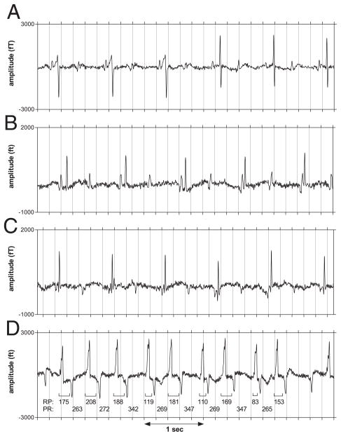
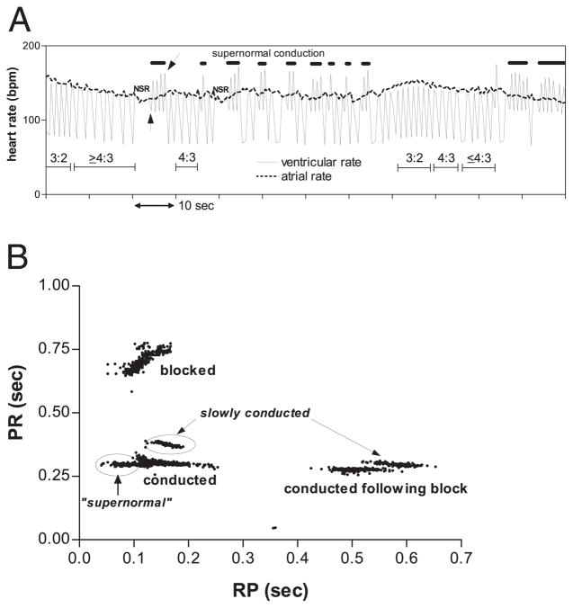
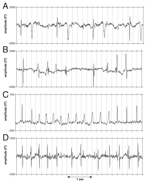
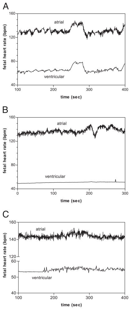
Results: Fetuses with 2 degrees AVB and isolated 3 degrees AVB showed: 1) diverse atrial rhythms and mechanisms of atrioventricular conduction during 2 degrees AVB; 2) junctional ectopic tachycardia and ventricular tachycardia during 3 degrees AVB; 3) reactive ventricular and atrial fetal heart rate (FHR) tracings at ventricular rates >56 beats/min; and 4) flat ventricular FHR tracings at ventricular rates <56 beats/min despite reactive atrial FHR tracings. In contrast, fetuses with 3 degrees AVB associated with structural cardiac disease exhibited predominantly nonreactive heart rate tracings and simpler rhythms.

Conclusions: Second-degree AVB, isolated 3 degrees AVB, and 3 degrees AVB associated with structural cardiac disease manifest distinctly different electrophysiological characteristics and outcome. Fetuses with 2 degrees AVB or isolated 3 degrees AVB commonly exhibited complex, changing heart rate and rhythm patterns; all 19 delivered fetuses are alive and healthy. Fetuses with structural cardiac disease and 3 degrees AVB exhibited largely monotonous heart rate and rhythm patterns and poor prognosis. Junctional ectopic tachycardia and/or ventricular tachycardia may be characteristic of an acute stage of heart block.


Figures

Figure 1
Figure 1. Rhythms in 2° Fetal AVB
Rhythm strips from second-degree (2°) atrioventricular block (AVB) subjects showing: (A) intermittent pre-excitation with QRS normalization in fetus #24 at 32 weeks’ gestation; (B) varying PR interval due to transient atrioventricular dissociation in fetus #24 at 37 weeks’ gestation; (C) period of low atrial rhythm (PR = 55 ms) in fetus #27 with 2 atrial rhythms at 24 weeks’ gestation; in the high rhythm (not shown), PR interval was approximately 107 ms; and (D) transition of sinus rhythm (1:1 conduction) from stable RP/PR to alternating RP/PR in fetus #25 at 33 weeks’ gestation. After several beats with relatively constant RP/PR, the fourth atrial beat conducts slowly, shortening RP of the succeeding beat. Rather than blocking, the fifth atrial beat not only conducts, it does so more rapidly than the prior beat, prolonging RP of the succeeding beat. The alternating (short–long) RP intervals of the ensuing beats continue to show PR prolongation during the “long” phase of the cycle and conduction at short RP during the “short” phase until block occurs, notably, during the “long” phase. The tracings were processed to remove maternal interference but were not signal averaged.
Figure 2
Figure 2. Fetal Heart Rate Patterns and AV Conduction Curves in Supernormal Conduction
(A) Ventricular and atrial heart rate tracing from last session of fetus #25, showing heart rate pattern changes associated with rhythm transitions. Wenckebach periods, indicated by the thin horizontal bars with conduction ratios above the bars, result in alternation of instantaneous ventricular heart rate between the atrial rate and half the atrial rate. A second pattern of alternation (thick horizontal lines) is due to beat-to-beat alternation of RP and PR, compatible with “supernormal” conduction. The RP and PR interval changes show a paradoxical positive correlation (Fig. 1D), which results in prominent beat-to-beat RR oscillations distinct from those due to Wenckebach periodicity. Usually, this pattern is immediately preceded by several or more beats of 1:1 sinus rhythm with constant RP/PR. The episodes initiate with a long RR interval (upward pointing arrow) and terminate with a short RR interval (slanted arrow) followed by a very long RR interval due to block of the following beat, as exemplified in the rhythm strip in Figure 1D. (B) Scatter plot of PR versus RP for each atrial beat in a 10-min recording during the last session of fetus #25. Atrial beats with RP >170 ms were always conducted, and the probability of conduction varied inversely with RP for 100 ms <RP <170 ms. During episodes of PR alternation (Fig. 1D); however, the data points formed 2 distinct clusters, enclosed by the ovals. One cluster consisted of beats conducted with long PR (slowly conducted); the other consisted of beats that conducted with short RP (“supernormal”). During episodes of PR alternation, beats with RP <100 ms were always conducted. Notice that in the ranges 100 ms <RP <250 ms and 450 ms <RP <650 ms, the conducted beats exhibited 2 distinct PR levels, suggesting the existence of 2 atrioventricular (AV) nodal pathways.
Figure 3
Figure 3. Ectopy and Tachycardia in 3° Fetal AVB
Rhythm strips from third-degree (3°) atrioventricular block (AVB) fetuses showing: (A) ventricular bigeminy with wide ectopic complexes; (B and C) episodes of nonsustained VT in fetuses #4 and #3, respectively; and (D) junctional ectopic tachycardia in fetus #14 with highly irregular ventricular rhythm at 19 weeks’ gestation.
Figure 4
Figure 4. Fetal Heart Rate Patterns in Fetal AVB
Atrial and ventricular fetal heart rate tracings from fetus #4 showing (A) reactive pattern at 28 weeks’ gestation and (B) nonreactive pattern at 32 weeks’ gestation. In the reactive pattern, the atrial and ventricular tracings were highly correlated; even variations as small as 3 beats/min (bpm) were often correlated. The degree of ventricular reactivity and the amplitude of the beat-to-beat fetal heart rate variability varied strongly with mean ventricular rate. At rates >70 beats/min, ventricular reactivity was exaggerated and the amplitude of the ventricular accelerations actually exceeded that of the atrial accelerations. As mean ventricular rate declined, the amplitude of the ventricular accelerations and the beat-to-beat fetal heart rate variability progressively decreased. Notice that atrial reactivity is present in the nonreactive pattern. Both patterns showed prominent atrial beat-to-beat variability due to the presence of ventriculophasic sinus arrhythmia. Unlike normal beat-to-beat variability, its amplitude does not diminish during heart rate acceleration. (C) Atrial and ventricular fetal heart rate tracings from fetus #3 at 30 weeks’ gestation showing abrupt transition from the nonreactive to the reactive pattern. AVB = atrioventricular block.

Comment in

References

    1. Menendez T, Achenbach S, Beinder E, et al. Usefulness of magnetocardiography for the investigation of fetal arrhythmias. Am J Cardiol. 2001;88:334–6. - PubMed
    1. Van Leeuwen P, Hailer B, Bader W, Geissler J, Trowitzsch E, Gronemeyer D. Magnetocardiography in the diagnosis of fetal arrhythmia. Br J Obstet Gynaecol. 1999;11:1200–8. - PubMed
    1. Wakai RT, Leuthold AC, Cripe L, Martin CB. Assessment of fetal rhythm in complete congenital heart block by magnetocardiography. Pacing Clin Electrophysiol. 2000;23:1047–50. - PubMed
    1. Zhao H, Wakai RT. Simultaneity of foetal heart rate acceleration and foetal trunk movement determined by foetal magnetocardiogram actocardiography. Phys Med Biol. 2002;47:839–46. - PubMed
    1. Leuthold A, Wakai RT, Martin CB. Noninvasive in utero assessment of PR and QRS intervals from the fetal magnetocardiogram. Early Hum Dev. 1999;54:235–43. - PubMed

Publication types