. 2008 Mar 28;283(13):8555-63.
doi: 10.1074/jbc.M710608200. Epub 2008 Jan 3.

Inhibitory role of Plk1 in the regulation of p73-dependent apoptosis through physical interaction and phosphorylation

Affiliations

Inhibitory role of Plk1 in the regulation of p73-dependent apoptosis through physical interaction and phosphorylation

Nami Koida et al. J Biol Chem. .

Abstract

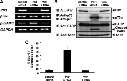
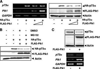
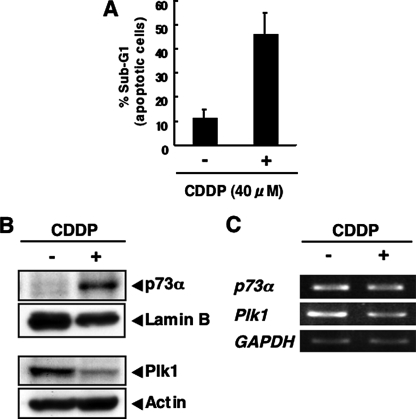
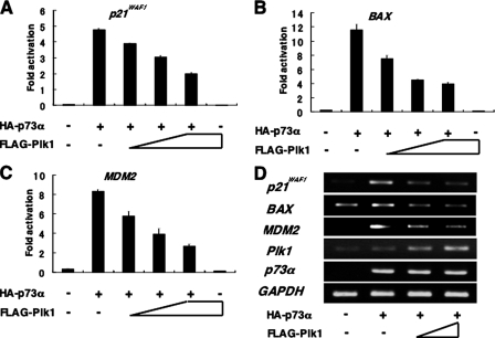
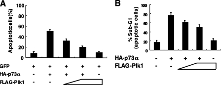
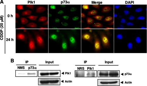
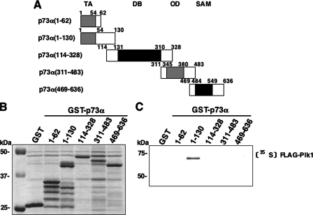
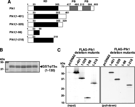
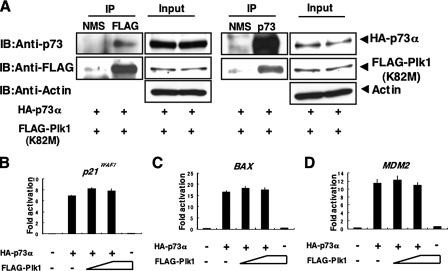
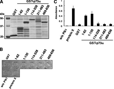
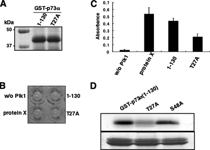
In response to DNA damage, p73 plays a critical role in cell fate determination. In this study, we have found that Plk1 (polo-like kinase 1) associates with p73, phosphorylates p73 at Thr-27, and thereby inhibits its pro-apoptotic activity. During cisplatin-mediated apoptosis in COS7 cells in which the endogenous p53 is inactivated by SV40 large T antigen, p73 was induced to accumulate in association with a significant down-regulation of Plk1. Consistent with these observations, Plk1 reduced the stability of the endogenous p73. Immunoprecipitation and in vitro pulldown assay demonstrated that p73 binds to the kinase domain of Plk1 through its NH(2)-terminal region. Luciferase reporter assay and reverse transcription-PCR analysis revealed that Plk1 is able to block the p73-mediated transcriptional activation. Of note, kinase-deficient Plk1 mutant (Plk1(K82M)) retained an ability to interact with p73; however, it failed to inactivate the p73-mediated transcriptional activation, suggesting that kinase activity of Plk1 is required for the inhibition of p73. Indeed, in vitro kinase assay indicated that p73 is phosphorylated at Thr-27 by Plk1. Furthermore, small interference RNA-mediated knockdown of the endogenous Plk1 in p53-deficient H1299 cells resulted in a significant increase in the number of cells with sub-G(1) DNA content accompanied by the up-regulation of p73 and pro-apoptotic p53(AIP1) as well as the proteolytic cleavage of poly(ADP-ribose) polymerase. Thus, our present results suggest that Plk1-mediated dysfunction of p73 is one of the novel molecular mechanisms to inhibit the p53-independent apoptosis, and the inhibition of Plk1 might provide an attractive therapeutic strategy for cancer treatment.


Figures

FIGURE 1.
FIGURE 1.
siRNA-mediated knockdown of Plk1 induces apoptosis in p53-deficient H1299 cells. A, siRNA-mediated knockdown of the endogenous Plk1. H1299 cells were transfected with control siRNA or with Plk1 siRNA (Plk1 siRNA). Forty-eight hours after transfection, total RNA was prepared and subjected to RT-PCR to examine the expression levels of Plk1, p73α, and p53AIP1. GAPDH was used as an internal control. B, immunoblotting analysis. H1299 cells were transfected as in A. Forty-eight hours after transfection, whole cell lysates were prepared and processed for immunoblotting (IB) with the indicated antibodies. For the detection of the endogenous p73α, whole cell lysates were subjected to immunoprecipitation (IP) with anti-p73 antibody followed by IB with anti-p73 antibody. Actin expression served as a control for equal loading of proteins in each lane. C, FACS analysis. H1299 cells were transfected as in A. Forty-eight hours after transfection, attached and floating cells were harvested, stained with PI, and their cell cycle distributions examined by flow cytometry.
FIGURE 2.
FIGURE 2.
Plk1 promotes proteolytic degradation of p73 in a proteasome-independent manner. A, enforced expression of Plk1 reduces the expression levels of p73. H1299 cells were co-transfected with the constant amount of HA-p73α (0.5 μg) expression plasmid together with or without the expression plasmid for FLAG-Plk1 (0.5 and 1.0 μg). Forty-eight hours after transfection, total RNA and whole cell lysates were prepared and subjected to RT-PCR (left panels) and IB with the indicated antibodies (right panels), respectively. B, H1299 cells were transfected with the expression plasmid for HA-p73α alone or with HA-p73α (0.5 μg) plus FLAG-Plk1 (0.5 μg) expression plasmids. Forty-eight hours after transfection, cells were treated with DMSO or with 20 μm of MG-132 for 6 h. Whole cell lysates were then extracted and subjected to IB with the indicated antibodies. C, H1299 cells were transfected with pcDNA3 or with FLAG-Plk1 expression plasmid. Forty-eight hours after transfection, whole cell lysates and total RNA were prepared and subjected to IP with anti-p73 antibody followed by IB with anti-p73 antibody (upper panels) and RT-PCR (lower panels), respectively. Actin was used as a loading control, and GAPDH was used as an internal control.
FIGURE 3.
FIGURE 3.
Inverse relationship between the endogenous expression levels of p73 and Plk1 in response to CDDP. A–C, COS7 cells were treated with 40 μm of CDDP or left untreated. Forty-eight hours after the treatment, floating and attached cells were collected, stained with PI, and their cell cycle distributions analyzed by flow cytometry (A). COS7 cells were treated with 40 μm of CDDP or left untreated. Forty-eight hours after the treatment, nuclear (upper panels) and whole cell lysates (lower panels) were prepared and subjected to IB with the indicated antibodies (B). Total RNA was also prepared and analyzed by RT-PCR (C).
FIGURE 4.
FIGURE 4.
Plk1 represses the p73-mediated transcriptional activation. A–C, H1299 cells (5 × 10⁴ cells) were co-transfected with the constant amount of HA-p73α expression plasmid (25 ng), 100 ng of p53/p73-responsive luciferase reporter construct bearing p21WAF1 (A), BAX (B), or MDM2 (C) promoter and 10 ng of Renilla luciferase reporter plasmid (pRL-TK) in the presence or absence of the increasing amounts of FLAG-Plk1 expression plasmid (50, 100, and 200 ng). To standardize the amounts of plasmid DNA per transfection, pcDNA3 was added to yield a total of 510 ng of plasmid. Forty-eight hours after transfection, cells were lysed, and their luciferase activities were measured. Data were normalized and presented as mean values ± S.D. of three independent experiments. D, RT-PCR analysis. H1299 cells were co-transfected with the constant amount of HA-p73α together with or without the increasing amounts of FLAG-Plk1 expression plasmid. Forty-eight hours after transfection, total RNA was prepared and analyzed for the expression levels of p21WAF1, BAX, and MDM2 by RT-PCR. Amplification of GAPDH serves as an internal control.
FIGURE 5.
FIGURE 5.
Plk1 inhibits the pro-apoptotic activity of p73. A, apoptotic assay. H1299 cells were seeded at a density of 2 × 10⁵ cells/6-well tissue culture plate and allowed to attach overnight. Next day, cells were co-transfected with the constant amount of GFP (100 ng) and HA-p73α (900 ng) expression plasmids together with or without the increasing amounts of FLAG-Plk1 expression plasmid (500 and 1000 ng). Total amount of plasmid DNA was kept constant (2 μg) with pcDNA3. Forty-eight hours after transfection, cells were fixe, and cell nuclei were stained with DAPI. The percentages of GFP-positive cells with apoptotic nuclei were plotted. B, FACS analysis. H1299 cells were co-transfected with the constant amount of the expression plasmid encoding HA-p73α (250 ng) together with or without the increasing amounts of FLAG-Plk1 expression plasmid (100 or 200 ng). Forty-eight hours after transfection, attached and floating cells were collected, stained with PI, and their cell cycle distributions analyzed by flow cytometry.
FIGURE 6.
FIGURE 6.
Interaction between Plk1 and p73 in cells. A, nuclear co-localization of Plk1 with p73 in response to CDDP. HeLa cells were treated with 20 μm CDDP (lower panels) or left untreated (upper panels). Twenty-four hours after the treatment cells were simultaneously probed with polyclonal anti-p73 antibody and monoclonal anti-Plk1 antibody for 1 h at room temperature. After extensive washing in PBS, cells were incubated with rhodamine-conjugated anti-mouse IgG (red) and fluorescein isothiocyanate-conjugated anti-rabbit IgG (green). Cell nuclei were stained with DAPI (blue). Merged images indicate the nuclear co-localization of Plk1 with p73α (yellow). B, immunoprecipitation. HeLa cells were exposed to 20 μm CDDP. Twenty-four hours after CDDP treatment, whole cell lysates were IP with NMS or with anti-p73 antibody followed by IB with anti-Plk1 antibody (left panel). Input lysates were analyzed by IB with the indicated antibodies. Reciprocal experiments are shown in the right panels.
FIGURE 7.
FIGURE 7.
NH2-terminal small domain of p73 is required for the interaction with Plk1. A, domain structure of wild-type p73α and schematic representation of GST-tagged p73α deletion mutants. TA, transactivation domain; DB, DNA-binding domain; OD, oligomerization domain; SAM, sterileα-motif domain. Numbers indicate amino acid positions. B, GST and GST-p73α fusion proteins were purified by glutathione-Sepharose beads and separated by 10% SDS-PAGE followed by Coomassie Brilliant Blue staining. C, in vitro pulldown assay. Equal amount of radiolabeled FLAG-Plk1 was incubated with GST or with the indicated GST-p73α fusion proteins. After incubation, GST or GST-p73α fusion proteins were recovered by glutathione-Sepharose beads, and bound materials were resolved by 10% SDS-PAGE followed by autoradiography.
FIGURE 8.
FIGURE 8.
Kinase domain of Plk1 is essential for the interaction with p73. A, schematic drawing of wild-type Plk1 and its deletion mutants. KD, kinase domain; PB, polo-box domain. B, Coomassie Brilliant Blue staining of GST-p73α-(1–130) used for this study. C, in vitro pulldown assay. Equal amount of GST-p73α-(1–130) was incubated with radiolabeled FLAG-Plk1 deletion mutants (left panel). After incubation, GST-p73α-(1–130) was precipitated by glutathione-Sepharose beads, and bound materials were separated by SDS-PAGE followed by autoradiography.
FIGURE 9.
FIGURE 9.
Kinase activity of Plk1 is required for the inhibition of p73. A, Plk1(K82M) retains an ability to interact with p73 in cells. COS7 cells were transiently co-transfected with the expression plasmids for HA-p73α and FLAG-Plk1(K82M). Forty-eight hours after transfection, whole cell lysates were prepared and subjected to IP with NMS or with anti-FLAG antibody. The immunoprecipitates were analyzed by IB with anti-p73 (1st panel) or with anti-FLAG (2nd panel) antibody. Input lysates were processed for IB with the indicated antibodies. Right panels show the results of the reciprocal experiments. B–D, luciferase reporter assay. H1299 cells were transiently co-transfected with the constant amount of HA-p73α expression plasmid (25 ng), 100 ng of luciferase reporter construct carrying p53/p73-responsive element derived from p21WAF1 (B), Bax (C), or MDM2 (D) promoter and 10 ng of pRL-TK together with or without the increasing amounts of the expression plasmid for FLAG-Plk1(K82M) (50 and 100 ng). Forty-eight hours after transfection, cells were lysed and their luciferase activities determined. Firefly luminescence signal was normalized based on the Renilla luminescence signal. Results were shown as fold induction of the firefly luciferase activity compared with control cells transfected with the empty plasmid alone.
FIGURE 10.
FIGURE 10.
Plk1 has an ability to phosphorylate p73 at its NH2-terminal region in vitro. A, Coomassie Brilliant Blue staining of GST or GST-p73α fusion proteins used for in vitro kinase reaction. B, in vitro kinase assay. GST or the indicated GST-p73α deletion mutants bound to glutathione-Sepharose beads were washed with washing buffer and resuspended in 90 μl of kinase reaction buffer. Protein X, which was supplied by manufacturer, was used as a positive control. 10 μl of the active form of Plk1 were added to the reaction mixtures and incubated at 30 °C for 30 min. The reaction mixtures were washed with washing buffer and then incubated with 100 μl of polyclonal anti-phospho-Thr antibody at room temperature for 30 min followed by incubation with horseradish peroxidase-conjugated anti-rabbit IgG at room temperature for 30 min. After incubation, 100 μl of substrate reagent were added to the reaction mixtures and incubated at room temperature for 5 min. Yellow coloration indicates the Plk1-mediated phosphorylation at Thr residue. After the addition of 100 μl of stop solution, supernatant was transferred into 96-well tissue culture plate, and the absorbance reading was carried out at 450/540 nm using the microplate reader (C).
FIGURE 11.
FIGURE 11.
Thr-27 of p73 is phosphorylated by Plk1 in vitro. A, Coomassie Brilliant Blue staining of GST-p73α-(1–130) and the mutant form of GST-p73α-(1–130) termed T27A where Thr-27 was substituted to Ala. B, in vitro kinase assay. In vitro kinase reactions were performed using GST-p73α-(1–130) and T27A as described in Fig. 10B. C, absorbance reading of the in vitro kinase reactions. D, standard in vitro kinase assay. GST-p73α-(1–130), T27A, and S48A were incubated with the active form of Plk1 in the presence of [γ-32P]ATP. The reaction mixtures were separated by SDS-PAGE and subjected to autoradiography (top panel). Bottom panel showed the Coomassie Brilliant Blue staining of the GST-p73α fusion proteins.

Similar articles

Cited by

References

    1. Kaghad, M., Bonnet, H., Yang, A., Creancier, L., Biscan, J. C., Valent, A., Minty, A., Chalon, P., Lelias, J. M., Dumont, X., Ferrara, P., McKeon, F., and Caput, D. (1997) Cell 90 809-819 - PubMed
    1. Melino, G., De Laurenzi, V., and Vousden, K. H. (2002) Nat. Rev. Cancer 2 605-615 - PubMed
    1. Ozaki, T., and Nakagawara, A. (2005) Cancer Sci. 96 729-737 - PMC - PubMed
    1. Bourdon, J. C., Fernandes, K., Murray-Zmijewski, F., Liu, G., Diot, A., Xirodiamas, D. P., Saville, M. K., and Lane, D. P. (2005) Genes Dev. 19 2122-2137 - PMC - PubMed
    1. Stiewe, T., Zimmermann, S., Frilling, A., Esche, H., and Putzer, B. M. (2002) Cancer Res. 62 3598-3602 - PubMed

Publication types

MeSH terms