. 2008 Apr 18;283(16):10297-309.
doi: 10.1074/jbc.M708017200. Epub 2008 Jan 3.

A deletion mutant of vitronectin lacking the somatomedin B domain exhibits residual plasminogen activator inhibitor-1-binding activity

Affiliations

A deletion mutant of vitronectin lacking the somatomedin B domain exhibits residual plasminogen activator inhibitor-1-binding activity

Christine R Schar et al. J Biol Chem. .

Abstract

Vitronectin and plasminogen activator inhibitor-1 (PAI-1) are important physiological binding partners that work in concert to regulate cellular adhesion, migration, and fibrinolysis. The high affinity binding site for PAI-1 is located within the N-terminal somatomedin B domain of vitronectin; however, several studies have suggested a second PAI-1-binding site within vitronectin. To investigate this secondary site, a vitronectin mutant lacking the somatomedin B domain (rDeltasBVN) was engineered. The short deletion had no effect on heparin-binding, integrin-binding, or cellular adhesion. Binding to the urokinase receptor was completely abolished while PAI-1 binding was still observed, albeit with a lower affinity. Analytical ultracentrifugation on the PAI-1-vitronectin complex demonstrated that increasing NaCl concentration favors 1:1 versus 2:1 PAI-1-vitronectin complexes and hampers formation of higher order complexes, pointing to the contribution of charge-charge interactions for PAI-1 binding to the second site. Furthermore, fluorescence resonance energy transfer between differentially labeled PAI-1 molecules confirmed that two independent molecules of PAI-1 are capable of binding to vitronectin. These results support a model for the assembly of higher order PAI-1-vitronectin complexes via two distinct binding sites in both proteins.


Figures
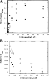
FIGURE 1.
FIGURE 1.
Recombinant forms of vitronectin bind to heparin. In a direct heparin-binding assay (A), a fixed concentration of heparin was immobilized in microtiter wells and blocked with casein. Varying concentrations of rVN (▴), rΔsBVN (□), native vitronectin (♦), and multimeric vitronectin (○) were incubated in the heparin-bound wells for 1 h. After rinsing the wells, bound vitronectin was detected using polyclonal anti-vitronectin antibodies and a peroxidase-labeled secondary antibody. Neutralization of the anticoagulant activity of heparin is followed in B. Heparin activity was measured by a decrease in thrombin activity due to inhibition by antithrombin. Thrombin activity was continuously monitored over time by measuring the hydrolysis of the chromogenic substrate Chromozym-TH. Variations in the Kapp were measured at various concentrations of vitronectin. Kapp rates were determined using the IGOR pro software and fitting the data using least-squares analysis. Kapp values were standardized to the reaction rate of heparin-catalyzed inhibition in the absence of vitronectin. Data points are shown at varying concentrations of multimeric vitronectin (○), rVN (▴), and rΔsBVN (□).
FIGURE 2.
FIGURE 2.
Recombinant ΔsBVN binds integrins, but not uPAR. The recombinant ΔsBVN bound to immobilized integrin GPIIbIIIa (A) and supported the binding of rabbit smooth muscle cells (B). A, varying concentrations of recombinant vitronectin (▴) and recombinant ΔsBVN (□) were incubated for 1 h in GPIIbIIIa-coated wells. Wells were washed, and bound vitronectin was detected using a monoclonal anti-vitronectin antibody and a peroxidase-labeled secondary antibody. The binding of rabbit smooth muscle cells to vitronectin is shown in B. Equal molar amounts (8 nm) of multimeric vitronectin (solid bar), rVN (open bar), and rΔsBVN (shaded bar) were immobilized in the wells of a tissue culture plate. Following blocking of the wells with a solution of 3.5% BSA in PBS, rabbit smooth muscle cells were added and allowed to adhere for 1 h. The amount of cell binding was determined by following the conversion of ρ-nitrophenyl phosphate to ρ-nitrophenol by alkaline phosphatase on the cell at an absorbance of 405 nm. The binding of smooth muscle cells to blocked wells alone was negligible and was subtracted as background. The recombinant ΔsBVN does not bind to soluble uPAR (C) or support U937 cell binding (D). C, an equal molar concentration of multimeric vitronectin (solid bar), rΔsBVN (open bar), or rVN (shaded bar) was used to coat the wells of a microtiter plate. The wells were blocked with 3% BSA in PBS before incubation with soluble uPAR. The amount of bound uPAR after washing was determined using polyclonal anti-uPAR antibodies and a peroxidase-labeled secondary antibody. D, stimulated U937 cells were stained with calcein, resuspended in serum-free medium with 15 nm uPA, and added to the wells of a microtiter plate that had been coated with equal molar concentrations of multimeric vitronectin (solid bar), rΔsBVN (open bar), or rVN (shaded bar). Cell binding was determined by measuring the fluorescent intensity of the calcein-labeled cells after washing with PBS. U937 cell binding to BSA-blocked wells accounted for 4% (∼850,000 units) of the binding to multimeric vitronectin and was subtracted as background. In both panels, the percent binding was calculated relative to multimeric vitronectin.
FIGURE 3.
FIGURE 3.
The recombinant ΔsBVN retains PAI-1-binding activity. Equal molar concentrations of native vitronectin (♦), multimeric vitronectin (○), rVN (▴), and rΔsBVN (□) were tested for binding to wild-type PAI-1 (A) or the 14-1B mutant form of PAI-1 (B) in a competitive assay where native vitronectin is immobilized in the wells of a microtiter plate. A fixed concentration of PAI-1 was added to wells in the presence of increasing concentrations of competing vitronectin. Bound PAI-1 was detected using a polyclonal anti-PAI-1 antibody. Percent binding inhibition was calculated using the equation, [(AxA0)/A0]*100, where A0 is the absorbance in the absence of competing vitronectin, and Ax is the absorbance at a given concentration of vitronectin.
FIGURE 4.
FIGURE 4.
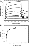
PAI-1 binds to the second site on vitronectin with lower affinity than the SMB domain. Representative sensorgrams obtained from the injection of varying concentrations of PAI-1 (1, 5, 10, 25, 50, 75, 250, and 500 nm) over immobilized rΔsBVN in a BIACORE 3000™ are shown in A. A duplicate series of sensorgram responses were normalized and plotted versus PAI-1 concentration (B). The points were fit to the 1:1 interaction model (Equation 1) to yield the KD of 29 ± 3 nm with an R2 value of 0.997.
FIGURE 5.
FIGURE 5.
FRET reveals the co-localization of two PAI-1 molecules on plasma vitronectin. Fluorescence donor PAI-1P1′-FL (20 nm) was reacted with vitronectin (20 nm) in the presence of “cold” wild-type PAI-1 (20 nm, dashed line) or with acceptor PAI-1P1′-TMR (20 nm, solid line) to yield a final 2:1 ratio (PAI-1-vitronectin). Emission spectra were recorded following excitation at 490 nm and corrected for dilution and the contribution of residual acceptor PAI-1P1′-TMR fluorescence when excited at 490 nm.
FIGURE 6.
FIGURE 6.
Sedimentation velocity analysis of equimolar mixtures of vitronectin and PAI-1 at varying NaCl concentrations. Mixtures of 3.2 μm vitronectin and PAI-1 were evaluated by analytical ultracentrifugation using the sedimentation velocity method. Data were analyzed using the c(s) method using SEDFIT (46). Plots showing the distribution of species in each sample in the range of sedimentation coefficients from 4 to 10 S are shown for samples in 20 mm sodium phosphate containing 150 mm (solid line), 250 mm (hatched line), and 500 mm (dashed line) NaCl.
FIGURE 7.
FIGURE 7.
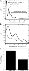
Analytical ultracentrifugation experiments demonstrate differences in higher order species formed by vitronectin with wild-type PAI-1 versus 14-1B PAI-1 variant. Mixtures of vitronectin (0.5 mg/ml) in a 1:1 molar ratio with wild-type PAI-1 or 14-1B PAI-1 were analyzed by sedimentation velocity (50,000 rpm) in a Beckman XL-I analytical ultracentrifuge. Data were analyzed using a continuous c(s) distribution model with the program SEDFIT (46). A, continuous c(s) distribution of complexes formed upon mixing of vitronectin with wild-type PAI-1. B, distribution profiles of higher order species formed between vitronectin and wild-type (solid line) or 14-1B stable mutant PAI-1 (dotted line). C, bar graph comparing the relative amounts of higher order species (s = 14–26) formed when vitronectin is mixed with either wild-type or 14-1B PAI-1. Peaks in the c(s) distribution in s = 14–26 range were integrated, and results were normalized relative to the total amount of protein loaded (concentrations are given in fringes). Error bars represent root mean square deviation values normalized to total loading concentration (fringes).

Similar articles

Cited by

References

    1. Holmes, R. (1967) J. Cell Biol. 32 297–308 - PMC - PubMed
    1. Preissner, K. T., and Muller-Berghaus, G. (1986) Eur. J. Biochem. 156 645–650 - PubMed
    1. Podor, T. J., Campbell, S., Chindemi, P., Foulon, D. M., Farrell, D. H., Walton, P. D., Weitz, J. I., and Peterson, C. B. (2002) J. Biol. Chem. 277 7520–7528 - PubMed
    1. Xu, D., Baburaj, K., Peterson, C. B., and Xu, Y. (2001) Proteins 44 312–320 - PubMed
    1. Mayasundari, A., Whittemore, N. A., Serpersu, E. H., and Peterson, C. B. (2004) J. Biol. Chem. 279 29359–29366 - PubMed

Publication types

LinkOut - more resources