. 2008 Mar;14(3):417-24.
doi: 10.1261/rna.874808. Epub 2008 Jan 3.

Loss of mir-146a function in hormone-refractory prostate cancer

Affiliations

Loss of mir-146a function in hormone-refractory prostate cancer

Shi-Lung Lin et al. RNA. 2008 Mar.

Abstract

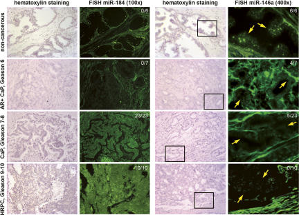
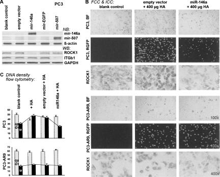
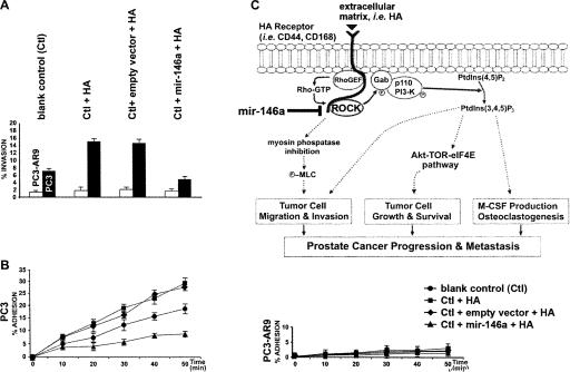
The pattern of microRNA (miRNA) expression is associated with the degree of tumor cell differentiation in human prostate cancer. MiRNAs bind complementarily to either oncogenes or tumor suppressor genes, which are consequently silenced, resulting in alterations of tumorigenecity. We have detected eight down-regulated and three up-regulated known miRNAs in androgen-independent human prostate cancer cells compared to those in androgen-dependent cells, using miRNA microarray analyses. These identified miRNAs showed the same expression patterns in hormone-refractory prostate carcinomas (HRPC) compared to androgen-sensitive noncancerous prostate epithelium as determined by fluorescent in situ hybridization assays in human prostate cancer tissue arrays. One of the eight down-regulated miRNAs, mir-146a, was selected and constitutively expressed to examine its effects on suppression of prostate cancer transformation from androgen-dependent to -independent cells as determined by in vitro tumorigenecity assays. Transfection of mir-146a, which perpetually express the miRNA, suppressed >82% of the expression of the targeted protein-coding gene, ROCK1, in androgen-independent PC3 cells, consequently markedly reducing cell proliferation, invasion, and metastasis to human bone marrow endothelial cell monolayers. Given that ROCK1 is one of the key kinases for the activation of hyaluronan (HA)-mediated HRPC transformation in vivo and in PC3 cells, mir-146a may function as a tumor-suppressor gene in modulating HA/ROCK1-mediated tumorigenecity in androgen-dependent prostate cancer.


Figures

FIGURE 1.
FIGURE 1.
MicroRNA microarray analyses of androgen-independent human prostate cancer cell lines, such as LNCaP C4-2B and PC3 (labeled by Cy3), compared to androgen-dependent cell lines, such as LNCaP and PC3-AR9 (labeled by Cy5), respectively (P <0.01, n = 3). Comparison of differentially expressed miRNAs in LNCaP versus C4-2B cells (upper panels) and PC3-AR9 versus PC3 cells (lower panels) shows 11 consistent miRNA alterations (white circles in the right two panels), including up-regulated mir-184, mir-361, and mir-424 (green dots) and down-regulated mir-19b, mir-29b, mir-128b, mir-146a, mir-146b, mir-221, mir-222, and mir-663 (red dots). Each of the four blue panels (the left and middle columns) represented the individual miRNA expression pattern in each cell line, showing miRNA expression from low abundant (blue–green) to high abundant (red) levels.
FIGURE 2.
FIGURE 2.
Fluorescent in situ hybridization analyses of mir-184 and mir-146a expression in human prostate cancer (CaP) tissue arrays in vivo. In view of prostatic epithelium, mir-184 expression was gradually increased consistent with cancer progression, while mir-146a expression (yellow arrows) was greatly diminished in most of the advanced prostatic carcinomas with Gleason scores over 7. Ratios (upper right corner) showed the number of positive samples versus the total patient numbers from each stage of prostate cancer. Simultaneous elevation of mir-184 and loss of mir-146a expression was observed in >75% of advanced prostate caner epithelium (P <0.001; n = 4), suggesting the significance of miRNA-mediated cancer transformation in vivo.
FIGURE 3.
FIGURE 3.
Mir-146a-mediated ROCK suppression in androgen-independent human prostate cancer PC3 cells. (A) Northern blotting (NB) of mir-146a and Western blotting (WB) of ROCK1 were concurrently performed. Transfection of mir-146a suppressed >82% of ROCK1 expression, while other off-target genes, such as integrin β1 (ITGb1) and GAPDH, were not affected (P <0.001; n = 4). (B) Immunocytochemical staining (ICC) of ROCK1 protein in fluorescent-labeled PC3-mir146a cells (FCC). Mir-146a-transfected PC3 cells coexpressed a fluorescent marker protein, RGFP (middle). The ROCK1 protein was markedly reduced in RGFP-positive PC3-mir146a cells, while the PC3 cells transfected with an empty vector showed elevated levels of active ROCK1 after HA stimulation. Cell proliferation rates were changed in response to these ROCK1 alterations, which displayed a much faster cell growth in HA-stimulated PC3 cells than those in PC3-mir146a cells, as shown in C. No significant change was found in PC3-AR9 cells with the same treatments. (C) Flow cytometry analysis of HA-stimulated cell proliferation (P <0.05; n = 4). Bar charts indicated the ratio of different cell populations (x axis) versus different treatments (y axis), including untreated control cells (Ctl), cells treated with 400 μg/mL HA (+ HA), cells treated with empty vector and then HA (empty vector+HA), and cells treated with mir-146a-expressing vector and then HA (mir-146a+HA). The white bar refers to the cell population resting in the G0/G1 nondividing phase, whereas the black bar represents the mitotic (M) cell population.
FIGURE 4.
FIGURE 4.
Effects of mir-146a-suppressed ROCK expression on HA-dependent cancer cell invasion and metastasis to hBMEC in androgen-independent PC3 and androgen-dependent PC3-AR9 cells. (A) Functional analysis of mir-146a-suppressed tumor invasion in matrigel chambers (P <0.05; n = 4). Black and white bars referred to PC3 and PC3-AR9 cells, respectively. In the presence of high HA density, a significant increase of tumor invasion was detected in PC3 cells (Ctl+HA), whereas mir-146a-mediated ROCK1 suppression in PC3-mir146a cells (Ctl+mir-146a+HA) completely reversed this tumorigenetic effect. Transfection of empty vector in PC3 cells (Ctl+empty vector+HA) did not provide the suppression effect on cancer cell invasion. No significant change was observed in PC3-AR9 cells with the treatment. (B) Comparison of cell adhesion to the hBMEC monolayer between PC3 and PC3-AR9 cells (P <0.05; n = 6). The x and y axes refer to time duration after treatments and cell population, respectively. Within 50 min, >29% of PC3 cell population adhered to the hBMEC monolayer after HA treatment (square mark) as compared with a 17% adhesion rate in PC3 cells without any treatment (circle mark). Mir-146a-mediated ROCK1 suppression in PC3-mir146a cells (triangle mark) greatly reduced the HA-stimulated cell adhesion rate from 29% to 7% (76% reduction). PC3-AR9 cells responded weakly to all treatments and barely showed any adhesion to hBMEC (right chart). (C) Schematic mechanisms by which mir-146a is involved in the HA-mediated HRPC transformation, including cancer cell proliferation (anti-apoptosis), invasion (migration), and metastasis to bone marrow tissues. In all three pathways, mir-146a, as a tumor suppressor gene, directly suppresses ROCK1 oncogene expression to prevent the tumorigenetic effects of HA-mediated ROCK signaling in prostate cancer.

References

    1. Bourguignon, L.Y.W., Singleton, P.A., Zho, H., Diedrich, F. Hyaluronan-mediated CD44 interaction with RhoGEF and Rho kinase promotes Grb2-associated binder-1 phosphorylation and phosphatidylinositol 3-kinase signaling leading to cytokine (macrophage-colony stimulating factor) production and breast tumor progression. J. Biol. Chem. 2003;278:29420–29434. - PubMed
    1. De Benedetti, A., Graff, J.R. eIF-4E expression and its role in malignancies and metastases. Oncogene. 2004;23:3189–3199. - PubMed
    1. Esquela-Kerscher, A., Slack, F.J. Oncomirs–MicroRNAs with a role in cancers. Nat. Rev. Cancer. 2006;6:259–269. - PubMed
    1. Grossmann, M.E., Huang, H., Tindall, D.J. Androgen receptor signaling in androgen-refractory prostate cancer. J. Natl. Cancer Inst. 2001;93:1687–1697. - PubMed
    1. Jagla, M., Feve, M., Kessler, P., Lapouge, G., Erdmann, E., Serra, S., Bergerat, J.P., Ceraline, J. A splicing variant of the androgen receptor detected in a metastatic prostate cancer exhibits exclusively cytoplasmic actions. Endocrinology. 2007;148:4334–4343. - PubMed

Publication types

MeSH terms