. 2008 Jan 15;105(2):775-80.
doi: 10.1073/pnas.0706447105. Epub 2008 Jan 3.

Homeostatic regulation of AMPA receptor expression at single hippocampal synapses

Affiliations

Homeostatic regulation of AMPA receptor expression at single hippocampal synapses

Qingming Hou et al. Proc Natl Acad Sci U S A. .

Erratum in

Abstract

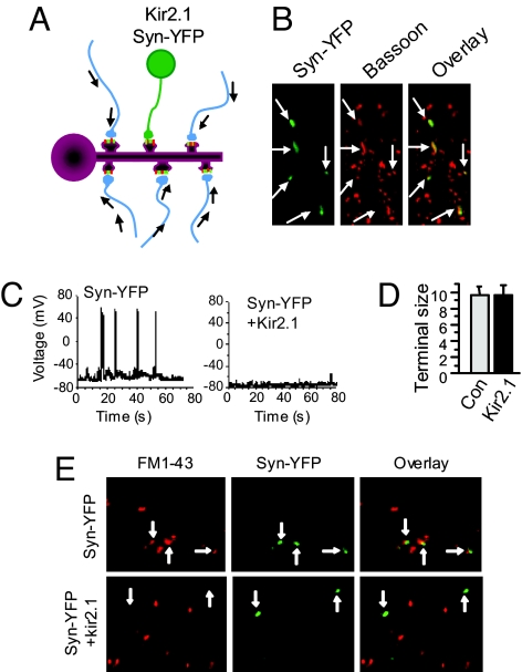
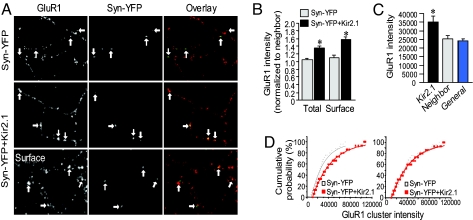
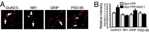
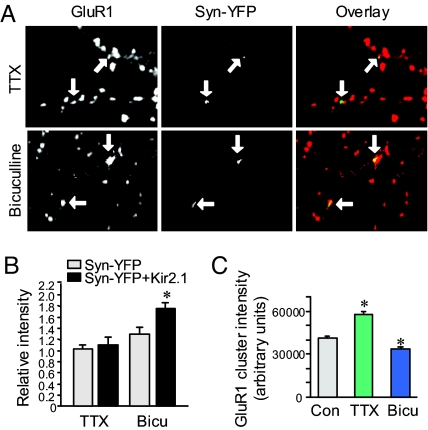
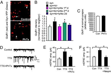
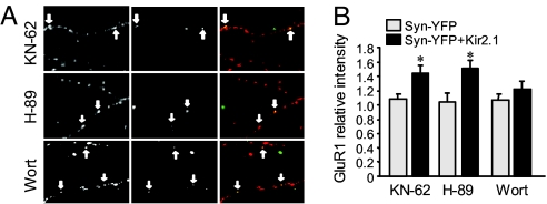
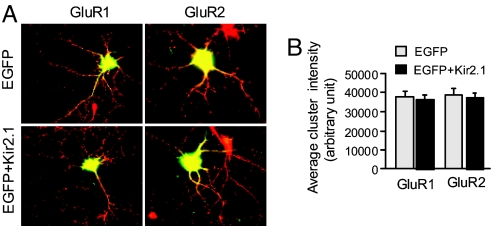
Homeostatic synaptic response is an important measure in confining neuronal activity within a narrow physiological range. Whether or not homeostatic plasticity demonstrates synapse specificity, a key feature characteristic of Hebbian-type plasticity, is largely unknown. Here, we report that in cultured hippocampal neurons, alpha-amino-3-hydroxy-5-methyl-isoxazole-4-propionic acid subtype glutamate receptor (AMPAR) accumulation is increased selectively in chronically inhibited single synapses, whereas the neighboring normal synapses remain unaffected. This synapse-specific homeostatic regulation depends on the disparity of synaptic activity and is mediated by GluR2-lacking AMPARs and PI3-kinase signaling. These results demonstrate the existence of synaptic specificity and the crucial role of AMPAR-gated calcium in homeostatic plasticity in central neurons.


Conflict of interest statement

The authors declare no conflict of interest.

Figures

Fig. 1.
Fig. 1.
Selective inhibition of individual synapses in cultured hippocampal neurons. (A) Schematic illustration of the experimental paradigm. Among a population of synapses on dendritic spines showing normal synaptic activity, one synapse, formed with a neuron expressing potassium channel Kir2.1, is inhibited. (B) In addition to the functional construct Kir2.1, neurons were cotransfected with YFP-tagged synapsin as a synapse marker. Immunostaining of the endogenous synaptic protein Bassoon showed a complete colocalization of synapsin-YFP and Bassoon (arrows). (C) Whole-cell recordings on transfected neurons. Under current-clamp configuration, small depolarizing synaptic activities were observed in both the Syn-YFP control neurons and the Kir2.1 neurons, with the amplitudes of the latter much smaller. (Left) In controls (n = 12), some depolarization pulses were big enough to trigger action potential (8–15 firings per min). (Right) In contrast, Kir2.1 neurons had hyperpolarized resting potentials and were mostly silent. (D) Axon terminal areas were estimated by measuring the YFP signals of synapsin-YFP. No changes in axon terminal size were found from neurons expressing synapsin-YFP (Con) and synapsin-YFP+Kir2.1 (Kir2.1) (Syn-YFP, 9.6 ± 1.1, n = 65; Kir2.1, 9.6 ± 1.2, n = 74). (E) Kir2.1 neuron terminals show reduced synaptic vesicle turnover. Two days after transfection with synapsin-YFP or together with Kir2.1, cells were rinsed twice with ACSF and incubated with 20 μM fixable FM1–43 in ACSF under basal conditions for 5–8 min. After three washes for 10 min with dye-free ACSF (containing no calcium to minimize spontaneous exocytosis), cells were fixed and imaged. Terminals from neurons expressing synapsin-YFP only (green) were loaded with FM dye (red, Upper), but those from Kir2.1-expressing neurons were largely lacking FM labeling (Lower), indicating a reduction in synaptic terminal activity. Arrows indicate colocalization of FM1-43 and Syn-YFP.
Fig. 2.
Fig. 2.
Homeostatic increase of GluR1 expression at inhibited single synapses. (A) GluR1 subunits were immunolabeled under permeant (Top and Middle) and nonpermeant (Bottom) conditions. At Kir2.1 synapses indicated by synapsin-YFP fluorescence (green), GluR1 (red) intensity was higher compared with neighboring normal synapses (Middle and Bottom), whereas GluR1 at synapsin-YFP-only synapses was not changed (Top). Arrows indicate synapsin-YFP terminals and the corresponding GluR1 puncta. (B) Pooled data of relative intensity of GluR1 stained under permeant (Total) and nonpermeant (Surface) conditions (Total: Syn-YFP, 1.03 ± 0.1, n = 42; Kir2.1, 1.3 ± 0.1, n = 48; P < 0.05. Surface: Syn-YFP, 1.1 ± 0.1, n = 54; Kir2.1, 1.6 ± 0.1, n = 54; P < 0.05, t test). (C) Absolute immunointensity of GluR1 clusters at Kir2.1 synapses (35,066 ± 3,136, n = 57), their neighbors (25,224 ± 1,609, n = 57) and the general synapse population (24,022 ± 960, n = 57) showed an increase at the inhibited but not the neighboring synapses, indicating that the increase in normalized GluR1 intensity was not due to a reduction in neighbor synapses. (D) Cumulative distribution of GluR1 puncta intensity. (Left) Data are fitted with single exponential. (Right) An overlap of the curves after multiplying a factor to the control (Syn-YFP) indicated a scaling up of AMPAR synaptic expression at Kir2.1 synapses. Error bars show SEM.
Fig. 3.
Fig. 3.
Homeostatic response is AMPAR-specific. (A) GluR2/3 expression, like GluR1, was increased at Kir2.1 synapses (Syn-YFP, 1.2 ± 0.1, n = 54; Kir2.1, 1.7 ± 0.1, n = 44; P < 0.05), but no changes were found for NMDAR subunit NR1 (Syn-YFP, 1.1 ± 0.1, n = 60; Kir2.1, 0.96 ± 0.1, n = 60; P > 0.05). Immunolabeling of the synaptic scaffolding protein PSD-95 and AMPAR-associated protein GRIP showed that at Kir2.1 synapses PSD-95 remained the same (Syn-YFP, 1.2 ± 0.1, n = 36; Kir2.1, 1.1 ± 0.1, n = 40; P > 0.05), whereas expression of GRIP increased (1.1 ± 0.1, n = 38 and 1.5 ± 0.1, n = 41 for Syn-YFP and Kir2.1 synapses, respectively; P < 0.05), suggesting a package delivery of AMPAR complexes during homeostatic response. (B) Pooled data.
Fig. 4.
Fig. 4.
Synapse-specific homeostatic response depends on disparity of synaptic strength. (A and B) When synaptic activity was equalized to low levels by application of TTX (1 μM) after Kir2.1 transfection, the homeostatic response in GluR1 expression was abolished (Syn-YFP, 1.10 ± 0.07, n = 52; Kir2.1, 1.18 ± 0.08, n = 54; P > 0.05). In contrast, when the network activity was enhanced by the GABAA receptor antagonist bicuculline (20 μM) to enlarge activity contrast, homeostatic increase in GluR1 expression at Kir2.1 synapses was enhanced (Syn-YFP, 1.30 ± 0.12, n = 40; Kir2.1, 1.76 ± 0.09, n = 43; P < 0.05). Arrows indicate Syn-YFP terminals and the corresponding GluR1 puncta. (C) The absolute intensity of synaptic GluR1 clusters of all synapses was increased by 2-d TTX treatment and was decreased by application of bicuculline, confirming the induction of global homeostatic synaptic regulation.
Fig. 5.
Fig. 5.
GluR2-lacking, calcium-permeable AMPARs are required for the induction of homeostatic response. (A) In neurons transfected with Syn-YFP and Kir2.1, homeostatic increase in GluR1 immunointensity (Upper) was completely blocked by coincubation with PhTx to specifically block the GluR2-lacking AMPARs (Lower). (B) Homeostatic response was blocked when PhTx was applied for only the first day or for 2 d after transfection. (Syn, 1.08 ± 0.06, n = 50; Kir2.1, 1.29 ± 0.08, n = 53; P < 0.05; Kir2.1 with PhTx-for 1 d, 1.09 ± 0.07, n = 61; P > 0.05; for 2 d 1.15 ± 0.06, n = 53; P > 0.05 compared with the Synapsin-YFP control). However, when supplemented only at the second day after transfection, PhTx failed to block the homeostatic response (1.24 ± 0.06, n = 56; P < 0.05 compared with Synapsin-YFP control), indicating a critical role of GluR2-lacking AMPARs in the early stages of homeostasis induction. (C) In neurons expressing Kir2.1 plus synapsin-YFP, 2-d incubation with PhTx did not change GluR1 intensity at normal synapses (n = 1,155). (D and E) TTX treatment (1 μM, 2 d) of cultured neurons induced global homeostatic increase in mEPSC amplitude (n = 15; P < 0.05). Coincubation of TTX (1 μM) and PhTx (5 μM) for 2 d abolished the TTX effect on mEPSC amplitude, indicating the critical role of GluR2-lacking AMPARs in homeostatic plasticity. A complete blockade of mEPSCs by CNQX confirmed AMPARs as the current mediators. (F) TTX-induced homeostatic increase in mEPSC amplitude was blocked by Naspm (n = 5).
Fig. 6.
Fig. 6.
Involvement of signaling kinases in homeostatic regulation. (A) Kinase inhibitors were applied to neurons after Kir2.1 transfection for 2 d. (B) Inhibition of PI3K activity (Wortmannin, 100 nM, n = 56) abolished the relative increase of GluR1 immunointensity at Kir2.1 synapses; whereas inhibition of CaMKII (KN-62, 10 μM, n = 47) and PKA (H-89, 1 μM, n = 57) did not affect the homeostatic response. Arrows indicate the inhibited synapses. *, P < 0.05, t test.
Fig. 7.
Fig. 7.
Postsynaptic suppression induces no homeostatic response in AMPAR synaptic expression. (A) Twelve-day cultured hippocampal neurons were transfected with EGFP as indicator or together with Kir2.1 to suppress neuronal excitability for 2 d. (B) Synaptic cluster intensity of GluR1 and GluR2 showed no difference compared with EGFP control, indicating that the homeostatic response in AMPAR expression is not sensitive to postsynaptic inhibition.

References

    1. Turrigiano GG, Nelson SB. Nat Rev Neurosci. 2004;5:97–107. - PubMed
    1. Davis GW. Annu Rev Neurosci. 2006;29:307–323. - PubMed
    1. O'Brien RJ, Kamboj S, Ehlers MD, Rosen KR, Fischbach GD, Huganir RL. Neuron. 1998;21:1067–1078. - PubMed
    1. Wierenga CJ, Ibata K, Turrigiano GG. J Neurosci. 2005;25:2895–2905. - PMC - PubMed
    1. Kilman V, van Rossum MC, Turrigiano GG. J Neurosci. 2002;22:1328–1337. - PMC - PubMed

Substances