. 2008 Mar;76(3):978-85.
doi: 10.1128/IAI.01354-07. Epub 2008 Jan 3.

A chemokine-degrading extracellular protease made by group A Streptococcus alters pathogenesis by enhancing evasion of the innate immune response

Affiliations

A chemokine-degrading extracellular protease made by group A Streptococcus alters pathogenesis by enhancing evasion of the innate immune response

Paul Sumby et al. Infect Immun. 2008 Mar.

Abstract

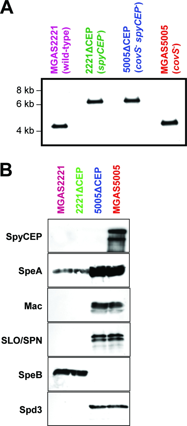
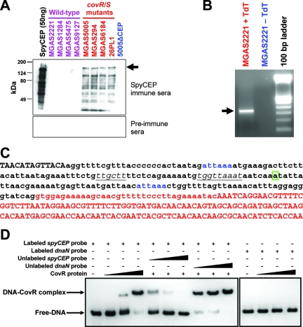
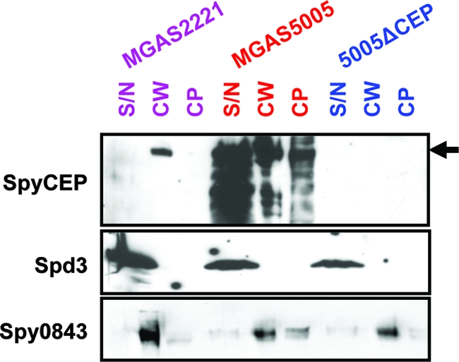
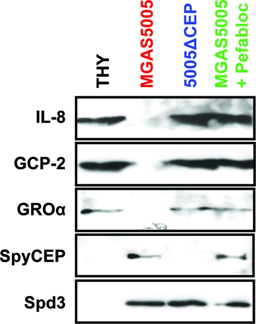
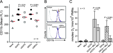
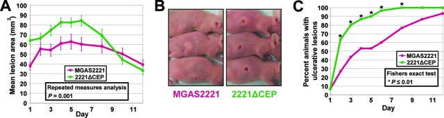
Circumvention of the host innate immune response is critical for bacterial pathogens to infect and cause disease. Here we demonstrate that the group A Streptococcus (GAS; Streptococcus pyogenes) protease SpyCEP (S. pyogenes cell envelope protease) cleaves granulocyte chemotactic protein 2 (GCP-2) and growth-related oncogene alpha (GROalpha), two potent chemokines made abundantly in human tonsils. Cleavage of GCP-2 and GROalpha by SpyCEP abrogated their abilities to prime neutrophils for activation, detrimentally altering the innate immune response. SpyCEP expression is negatively regulated by the signal transduction system CovR/S. Purified recombinant CovR bound the spyCEP gene promoter region in vitro, indicating direct regulation. Immunoreactive SpyCEP protein was present in the culture supernatants of covR/S mutant GAS strains but not in supernatants from wild-type strains. However, wild-type GAS strains do express SpyCEP, where it is localized to the cell wall. Strain MGAS2221, an organism representative of the highly virulent and globally disseminated M1T1 GAS clone, differed significantly from its isogenic spyCEP mutant derivative strain in a mouse soft tissue infection model. Interestingly, and in contrast to previous studies, the isogenic mutant strain generated lesions of larger size than those formed following infection with the parent strain. The data indicate that SpyCEP contributes to GAS virulence in a strain- and disease-dependent manner.


Figures

FIG. 1.
FIG. 1.
Confirmation of spyCEP mutations in the isogenic mutant strains 5005ΔCEP and 2221ΔCEP. (A) Southern blot confirming correct molecular construction of isogenic mutant strains 5005ΔCEP and 2221ΔCEP. Genomic DNA from parent GAS strains MGAS5005 and MGAS2221 and putative spyCEP mutant strains 5005ΔCEP and 2221ΔCEP was digested with NcoI, separated by agarose gel electrophoresis, transferred to a membrane, and probed with a region of DNA flanking spyCEP. The expected hybridizing fragment size for the parental strains is 4,205 bp, whereas the expected size for a spyCEP mutant strain is 6,674 bp. (B) Western immunoblots confirming the absence of immunoreactive SpyCEP protein in culture supernatants of isogenic mutant strain 5005ΔCEP. Protein was isolated from overnight culture supernatants of parent strains MGAS5005 and MGAS2221 and spyCEP mutant derivatives 5005ΔCEP and 2221ΔCEP. Reactivities with antibodies to SpyCEP, streptococcal pyrogenic exotoxin A (SpeA), SpeB, streptolysin O (SLO), S. pyogenes NAD-glycohydrolase (SPN), Mac-1-like protein (Mac), and S. pyogenes DNase 3 (Spd3) were determined. Different protein secretion patterns between MGAS5005 and MGAS2221 are attributed to a mutation within covS in strain MGAS5005 encoding the sensor kinase component of the virulence-regulating two-component system CovR/S (30).
FIG. 2.
FIG. 2.
spyCEP is regulated directly by the CovR/S two-component system. (A) Enhanced SpyCEP secretion by GAS isolates containing mutations in the two-component signal transduction system covR/S. Protein from 200-μl aliquots of overnight culture supernatants of wild-type (purple) or covR/S mutant (red) GAS strains were concentrated and separated by SDS-PAGE. All strains are clinical isolates with the exception of strain 26PL1, which is an isogenic covS mutant of parental strain MGAS2221 (30). Western immunoblots were probed with mouse preimmune serum or SpyCEP immune serum. Fifty-nanogram aliquots of purified recombinant SpyCEP (black) and supernatant protein from mutant strain 5005ΔCEP (blue) were loaded as positive and negative controls, respectively. The arrow indicates the full-size SpyCEP protein. (B) A single PCR product produced by 5′ RACE of the spyCEP gene. Poly(C)-tailed cDNA (+TdT) created by 5′ RACE gave a single PCR product following separation by agarose gel electrophoresis. The absence of amplification following use of untailed (-TdT) cDNA confirmed the specificity of the amplification reaction. (C) DNA sequence spanning the spyCEP promoter. The 5′ end of the spyCEP gene is labeled red. The transcriptional start site identified by 5′ RACE is boxed. The putative extended −10 and −35 promoter sequences are italicized and underlined. Putative CovR binding sites are blue. Lowercase bases indicate the 210-bp DNA fragment amplified for use in electrophoretic mobility shift assays. (D) CovR binds to the spyCEP promoter region. Electrophoretic mobility shift assays identified that purified CovR binds in a specific manner to the spyCEP promoter, such that CovR can be titrated away from the labeled spyCEP promoter DNA through addition of unlabeled spyCEP promoter DNA but not by addition of identical concentrations of an unrelated chromosomal region (the dnaN gene).
FIG. 3.
FIG. 3.
SpyCEP can be localized to the GAS cell wall. Fractions corresponding to cytoplasmic (CP), cell wall-anchored (CW), and secreted (SN) proteins were isolated for GAS strains MGAS2221 (wild type; purple), MGAS5005 (covS variant; red), and 5005ΔCEP (spyCEP mutant and covS variant; blue) and subjected to Western immunoblot analyses. All protein fractions from strain MGAS5005 had reactivity to the SpyCEP-specific antiserum. Reactivity to the SpyCEP antiserum was only observed in the cell wall protein fraction of strain MGAS2221 and was absent in all fractions from strain 5005ΔCEP. The secreted protein Spd3 and cell wall-anchored protein Spy0843 were probed as loading controls. Note that due to greater protein concentrations the S/N samples resulted in lanes wider than other lanes. The arrow indicates the full-size SpyCEP protein.
FIG. 4.
FIG. 4.
SpyCEP cleaves the human chemokines IL-8, GCP-2, and GROα. Recombinant chemokine proteins were incubated with filtered supernatants from strain MGAS5005 (red) and isogenic mutant 5005ΔCEP (blue) before use in Western immunoblot analyses. Supernatant from strain MGAS5005, but not from strain 5005ΔCEP, resulted in the degradation of IL-8, GCP-2, and GROα, as evidenced by the loss of reactivity to polyclonal antibodies. Addition of the SpyCEP inhibitor Pefabloc to MGAS5005 supernatant reaction mixtures inhibited chemokine degradation (green). Incubation of chemokines with sterile THY broth served as a negative control for cleavage (black). Spd3 reactivity was assayed as a control for loading.
FIG. 5.
FIG. 5.
SpyCEP inhibits priming of human neutrophils by GCP-2 and GROα. (A and B) Surface expression of CD11b. Neutrophils were cultured with 10 nM GCP-2 or GROα or those cleaved with SpyCEP (cGCP2 or cGROα). Results are the mean fluorescence (mean FL1) of neutrophils from four separate individuals (each symbol). The red line is the mean. Panel B is from a representative flow cytometry experiment. (C) SpyCEP inhibits priming for fMLF-stimulated neutrophil O2 generation. Human neutrophils were activated by 1 μM fMLF with or without priming by chemokines as indicated. Results are the means ± standard deviations of three separate experiments. Data were analyzed with a repeated-measures analysis of variance and Bonferroni's posttest for multiple comparisons.
FIG. 6.
FIG. 6.
SpyCEP contributes to M1T1 GAS mouse pathogenesis. (A) Soft tissue infection model. Data show the mean skin lesion area over time for groups of 30 mice infected subcutaneously with 1 × 10⁷ CFU of either strain MGAS2221 or 2221ΔCEP. Error bars indicate the standard errors of the means. (B) Photographs of representative mice 2 days after subcutaneous infection with strain MGAS2221 or spyCEP isogenic mutant 2221ΔCEP. (C) Mice infected subcutaneously with spyCEP isogenic mutant strain 2221ΔCEP develop ulcerative lesions at a significantly greater rate than mice infected with parental strain MGAS2221. Asterisks indicate time points of statistical significance.

Similar articles

Cited by

References

    1. Beres, S. B., and J. M. Musser. 2007. Contribution of exogenous genetic elements to the group A Streptococcus metagenome. PLoS ONE 2e800. - PMC - PubMed
    1. Biswas, I., P. Germon, K. McDade, and J. R. Scott. 2001. Generation and surface localization of intact M protein in Streptococcus pyogenes are dependent on sagA. Infect. Immun. 697029-7038. - PMC - PubMed
    1. Brinkmann, V., U. Reichard, C. Goosmann, B. Fauler, Y. Uhlemann, D. S. Weiss, Y. Weinrauch, and A. Zychlinsky. 2004. Neutrophil extracellular traps kill bacteria. Science 3031532-1535. - PubMed
    1. Carlsson, F., C. Sandin, and G. Lindahl. 2005. Human fibrinogen bound to Streptococcus pyogenes M protein inhibits complement deposition via the classical pathway. Mol. Microbiol. 5628-39. - PubMed
    1. Cockerill, F. R., III, R. L. Thompson, J. M. Musser, P. M. Schlievert, J. Talbot, K. E. Holley, W. S. Harmsen, D. M. Ilstrup, P. C. Kohner, M. H. Kim, B. Frankfort, J. M. Manahan, J. M. Steckelberg, F. Roberson, W. R. Wilson, et al. 1998. Molecular, serological, and clinical features of 16 consecutive cases of invasive streptococcal disease. Clin. Infect. Dis. 261448-1458. - PubMed

Publication types

MeSH terms