. 2008 May 15;111(10):5215-22.
doi: 10.1182/blood-2007-09-113092. Epub 2008 Jan 3.

Cell cycle-dependent chromatin loading of the Fanconi anemia core complex by FANCM/FAAP24

Affiliations

Cell cycle-dependent chromatin loading of the Fanconi anemia core complex by FANCM/FAAP24

Jung Min Kim et al. Blood. .

Abstract

Fanconi anemia (FA) is a genetic disease characterized by congenital abnormalities, bone marrow failure, and cancer susceptibility. A total of 13 FA proteins are involved in regulating genome surveillance and chromosomal stability. The FA core complex, consisting of 8 FA proteins (A/B/C/E/F/G/L/M), is essential for the monoubiquitination of FANCD2 and FANCI. FANCM is a human ortholog of the archaeal DNA repair protein Hef, and it contains a DEAH helicase and a nuclease domain. Here, we examined the effect of FANCM expression on the integrity and localization of the FA core complex. FANCM was exclusively localized to chromatin fractions and underwent cell cycle-dependent phosphorylation and dephosphorylation. FANCM-depleted HeLa cells had an intact FA core complex but were defective in chromatin localization of the complex. Moreover, depletion of the FANCM binding partner, FAAP24, disrupted the chromatin association of FANCM and destabilized FANCM, leading to defective recruitment of the FA core complex to chromatin. Our results suggest that FANCM is an anchor required for recruitment of the FA core complex to chromatin, and that the FANCM/FAAP24 interaction is essential for this chromatin-loading activity. Dysregulated loading of the FA core complex accounts, at least in part, for the characteristic cellular and developmental abnormalities in FA.


Figures

Figure 1
Figure 1
FANCM is detected exclusively in the chromatin-containing fraction. (A) Experimental protocol for cell fractionation described in this study. (B) HeLa cells were treated with MMC (50 ng/mL for 17 hours) and HU (2 mM for 17 hours) and then fractionated into the cytosol and nucleoplasmic fraction (S) and the chromatin-containing fraction (P). (C) HeLa cells were fractionated into S and P as in Figure 1A. The pellet was further extracted with 1% Triton X-100 (lanes 5-8) or 250 mM NaCl (lanes 9-12). After centrifugation, the supernatant and pellet were analyzed by Western blotting. (D) Cell extracts were incubated with lambda phosphatase and immunoblotted for the indicated proteins.
Figure 2
Figure 2
FANCM undergoes cell cycle–dependent phosphorylation. (A) Asynchronously growing cells (lane 1), cells arrested in G1/S phase with double thymidine blocking and released into S phase (lanes 2-7), or cells arrested in metaphase with nocodazole and released into G1 phase (lanes 8-12) were fractionated into S and P and immunoblotted for the indicated proteins. Asyn indicates asynchronous cells. Synchrony was monitored by flow cytometric analysis. The SDS-PAGE gel used here was 4% Tris-glycine. (B) Experimental protocol used to prepare cell extracts described in panels C and D. (C) Either exponentially growing Flag-FANCL HeLa cells (Asyn; lanes 1-4), cells released from double thymidine block for 3 hours (S phase; lanes 5-8), or cells arrested in mitotic phase with nocodazole (M phase; lanes 9-12) were fractionated into S100 and S300. As a negative control for immunoprecipitation with FLAG antibody, parental HeLa cells (lanes 13-16) were used. Each fraction was immunoprecipitated with FLAG-M2 agarose beads, and the precipitates were immunoblotted for the indicated proteins. (D) Cells were arrested in the G1/S phase by double thymidine block (lanes 1-4) and released into S phase (lanes 5-16). Alternatively, cells were arrested in mitotic phase with nocodazole (laness 17-20) and fractionated into S100 and S300. Proteins from each fraction were immunoprecipitated with FLAG-M2 agarose beads, and the precipitates were immunoblotted with the indicated antibodies. Note that for the Western blots of Flag-FANCL (fourth row), immunoprecipitation samples were exposed for a shorter time than for input samples. This is indicated by the vertical lines in between. This was necessitated by the fact that the intensity of IgG bands that appeared on immunoprecipitation samples was substantially greater than that of the input Flag-FANCL bands. Entire images come from the identical gel.
Figure 3
Figure 3
The chromatin association of the FA core complex is impaired in FANCM-depleted cells. (A) EUFA867 lymphoblast and wild-type cells (GM02254A.L) were treated with MMC (150 ng/mL for 17 hours) and fractionated into S and P. α-tubulin and histone H3 are loading controls for S and P fractions, respectively. (B) HeLa cells transfected with the indicated siRNAs were analyzed by immunoblotting to insure FANCM or FANCA protein knockdown. (C) HeLa cells treated with the indicated siRNAs for 72 hours were treated with MMC (50 ng/mL for 17 hours). The whole-cell extracts were immunoblotted for the indicated proteins. The α-tubulin is a loading control for S fraction, and MCM3 was used as a loading control for P fraction. The crossreactive band are labeled with asterisks in panels A and C. (D) HeLa cells transfected with indicated siRNA for 72 hours were incubated without or with MMC (50 ng/mL for 17 hours) and then stained with anti-FANCG antibody, either without or with 0.3% Triton X-100 pre-extraction before fixation.
Figure 4
Figure 4
FANCM is not essential for the assembly of the FA core complex. (A) Parental HeLa cells (lane 1) and Flag-FANCL HeLa cells (lanes 2,3) transfected with indicated siRNAs for 72 hours were incubated with MMC (50 ng/mL for 17 hours) and harvested and immunoblotted for the indicted protein. (B) Cells treated as in panel A were extracted with 300 mM NaCl-CSK buffer and immunoprecipitated with FLAG-M2 agarose beads. The precipitates (lanes 1-3) and input (lanes 4-6) were immunoblotted for the indicated proteins.
Figure 5
Figure 5
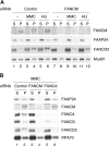
The chromatin association of FAAP24 depends on FANCM. (A) HeLa cells transfected with control or FANCM siRNA for 72 hours were treated with MMC (50 ng/mL for 17 hours) or HU (2 mM for 17 hours) and harvested. Fractionated extracts were immunoblotted for the indicated proteins. Mus81 was used a loading control for both fractions. (B) HeLa cells transfected with the indicated siRNAs for 72 hours were treated with MMC (50 ng/mL for 17 hours) and harvested. Fractionated extracts were immunoblotted for the indicated proteins. RPA70 was used a loading control for both fractions.
Figure 6
Figure 6
FANCM requires FAAP24 for its stable association in chromatin. (A) HeLa cells were transfected with indicated siRNAs for 72 hours, and then whole-cell extracts were immunoblotted. (B) HeLa cells transfected with indicated siRNAs for 72 hours were treated with MMC (50 ng/mL for 17 hours) or HU (2 mM for 17 hours), and whole-cell extracts were immunoblotted for the indicated proteins. (C) HeLa cells transfected with indicated siRNAs for 72 hours, and then incubated with MMC (50 ng/mL) for 7 hours, 12 hours, and 20 hours. Cells were then extracted and fractionated, and each fraction was immunoblotted for the indicated proteins.
Figure 7
Figure 7
Model for the role of FANCM in regulating the FA pathway. (A) HeLa cells silenced for the indicated proteins by siRNA (columns) were analyzed for MMC sensitivity, FANCD2 monoubiquitination, presence of FA core complex, and chromatin localization of FA core complex (rows). + indicates indistinguishable from control siRNA–treated cells; and −, defective. (B) Schematic model describing the function of FANCM in the chromatin recruitment of the FA core complex. The FANCM-FAAP24 complex associates with chromatin throughout the cell cycle. Early in the cell cycle (G1 phase), the FA core (A/B/C/E/F/G/L complex) is assembled but does not associate with FANCM-FAAP24 complex in chromatin. In S phase, phosphorylated FANCM can recruit the FA core complex to chromatin, possibly to replication forks, and induce E3 ubiquitin ligase activity, resulting in monoubiquitination of FANCD2 and FANCI. In G2/M phase, hyperphosphorylated FANCM may promote the release of the FA core complex, and USP1 may deubiquitinate FANCD2 and FANCI monoubiquitination.

Comment in

References

    1. Grompe M, van de Vrugt H. The Fanconi family adds a fraternal twin. Dev Cell. 2007;12:661–662. - PubMed
    1. Medhurst AL, Huber PA, Waisfisz Q, de Winter JP, Mathew CG. Direct interactions of the five known Fanconi anaemia proteins suggest a common functional pathway. Hum Mol Genet. 2001;10:423–429. - PubMed
    1. Medhurst AL, Laghmani el H, Steltenpool J, et al. Evidence for subcomplexes in the Fanconi anemia pathway. Blood. 2006;108:2072–2080. - PMC - PubMed
    1. Meetei AR, de Winter JP, Medhurst AL, et al. A novel ubiquitin ligase is deficient in Fanconi anemia. Nat Genet. 2003;35:165–170. - PubMed
    1. Meetei AR, Medhurst AL, Ling C, et al. A human ortholog of archaeal DNA repair protein Hef is defective in Fanconi anemia complementation group M. Nat Genet. 2005;37:958–963. - PMC - PubMed

Publication types